

Isotype Control Selection: A Practical Checklist for Researchers
Use an isotype control when you need to check for nonspecific binding or incomplete blocking — and match it on three attributes without exception: host species, immunoglobulin class and subclass (including clonality), and conjugate or fluorochrome. If you are setting gates in a multicolor flow panel, reach for a Fluorescence Minus One (FMO) control instead. Here is the minimum checklist before you order or run your assay:
- Match host species. A mouse IgG1 primary requires a mouse IgG1 isotype control, not a rat or rabbit equivalent.
- Match Ig class and subclass exactly. IgG1 and IgG2a are not interchangeable, even from the same species.
- Match clonality. Monoclonal primary antibody gets a monoclonal isotype control; polyclonal gets a polyclonal control.
- Match conjugate or fluorochrome. A PE-conjugated primary needs a PE-conjugated isotype at the same fluorochrome-to-protein (F:P) ratio when possible.
- Match concentration, not just volume. Run the isotype at the same microgram-per-milliliter concentration as your titrated primary, not the manufacturer’s suggested starting concentration.
- Consider alternatives first. For multicolor panels, FMO controls and unstained or negative-cell gating often provide more reliable gate placement than isotype controls alone.
Key Takeaways
Correct isotype control selection requires matching host species, Ig class and subclass, clonality, and conjugate to the primary antibody, then titrating to the same working concentration — while reserving FMO controls for gate-setting in multicolor panels.
| Point | Details |
|---|---|
| Match four attributes | Host species, Ig class/subclass, clonality, and conjugate must all match the primary antibody. |
| Always titrate | Run the isotype at the same microgram-per-milliliter concentration as your titrated primary, not the stock suggestion. |
| FMOs for gating | Use FMO controls to set gates in multicolor panels; isotype controls do not account for spillover and spreading error. |
| Isotypes have a narrow role | Use isotype controls to check Fc blocking or detect gross reagent artifacts, not as a routine gate-setting tool. |
| Mayflowerbio for reagents | Mayflowerbio’s antibody catalog includes matched isotype controls with lot-specific QC documentation available on request. |
Table of Contents
- How to choose the right isotype control for your assay
- Stepwise selection: what to match before you order
- How to titrate an isotype control and set up your control tubes
- Common mistakes and when isotype controls mislead you
- Better alternatives: FMOs, unstained controls, and reducing background
- Key protocol references and search terms for validated guidance
- The case for treating isotype controls as a diagnostic tool, not a default
- Mayflowerbio supports your antibody and reagent needs
- Sources
- FAQ
How to choose the right isotype control for your assay
An isotype control is an antibody of the same immunoglobulin class, subclass, and conjugation as your primary antibody, raised against an irrelevant target absent from your sample. Its job is to reveal signal that comes from nonspecific binding or Fc receptor engagement, not from genuine target recognition. That sounds straightforward, but the practical value of isotype controls depends heavily on the assay type and how carefully they are matched.
In flow cytometry, isotype controls remain useful in a narrow set of scenarios: verifying that Fc blocking is working, catching lot-specific reagent artifacts, and providing a rough estimate of background in single-color experiments on simple samples. In immunofluorescence (IF) and immunohistochemistry (IHC), they serve a similar role — detecting secondary antibody cross-reactivity or tissue autofluorescence that is not target-specific. Fixed and permeabilized samples in IF can show elevated nonspecific binding, and an isotype run in parallel helps distinguish that background from real intracellular signal.
That consensus view matters. For routine surface-marker immunophenotyping on well-characterized samples, an isotype control rarely tells you anything an unstained tube does not. The NCBI flow cytometry protocols reference describes the distinct roles of unstained, single-color, FMO, and isotype controls — and the takeaway is that each control answers a different question. Conflating them is where most errors begin.
Stepwise selection: what to match before you order
Isotype control selection is not a single decision — it is a short sequence of attribute checks. Work through them in order.
- Confirm host species. Check the primary antibody datasheet. Mouse, rat, rabbit, Armenian hamster, and goat are the most common hosts. Your isotype control must come from the same species.
- Confirm Ig class and subclass. Mouse IgG1, IgG2a, IgG2b, and IgG3 have different Fc receptor affinities. A mismatch here produces a control that does not replicate the primary’s nonspecific binding pattern.
- Confirm clonality. Monoclonal isotype controls are preferable for monoclonal primaries because they have a defined, consistent binding profile. Polyclonal isotype controls are appropriate for polyclonal primaries.
- Match conjugate and fluorochrome. The conjugate must be identical — same fluorochrome, same tandem dye formulation if applicable. Abcam and Bio-Rad both publish isotype control panels with matched conjugates; check their product pages for your fluorochrome.
- Check the F:P ratio. Fluorochrome-to-protein ratio affects brightness. A high-F:P isotype control will appear brighter than a low-F:P primary even at the same microgram concentration, inflating apparent background. When the vendor lists F:P on the datasheet, select an isotype with a ratio within the same range.
- Use the same buffer and matrix. Stain isotype controls in the same buffer (PBS/BSA, FACS buffer) and, where possible, the same sample matrix as the primary. Serum proteins in the matrix affect nonspecific binding.
- Run lot-specific QC checks. Different lots of the same isotype from the same vendor can behave differently, so confirm the lot number and request the vendor’s QC certificate. Abcam provides lot-specific datasheets; Bio-Rad’s antibody pages include lot release criteria.
- Consider an isoclonic control for critical experiments. An isoclonic control uses the same clone as the primary antibody but conjugated to a different fluorochrome or used at a non-binding concentration. It controls for clone-specific nonspecific binding more precisely than a generic isotype.
Pro Tip: Record the isotype control’s lot number, clone, F:P ratio, and titration curve in your lab notebook alongside the primary antibody entry. Journals increasingly require this level of documentation in methods sections, and having it ready saves revision rounds.
How to titrate an isotype control and set up your control tubes
Titration is non-negotiable. Running an isotype control at the manufacturer’s suggested concentration without titrating it against your primary is one of the most common sources of misleading data in published flow cytometry experiments.
Titration protocol:
- Prepare a serial dilution series of the isotype control (typically 1:2 or 1:4 steps) spanning at least four concentrations bracketing your expected working concentration.
- Stain replicate aliquots of your sample in the same staining buffer used for the primary antibody.
- Include a viability dye (e.g., DAPI, 7-AAD, or a fixable amine-reactive dye) in every tube. Dead cells bind antibodies nonspecifically and will inflate your isotype signal if not excluded.
- Add Fc block (e.g., human FcR blocking reagent for human samples, mouse Fc block for murine samples) before antibody addition, using the same blocking conditions as your primary staining.
- Acquire all tubes on the same instrument settings. Do not adjust voltages between the isotype and primary tubes.
- Identify the concentration at which the isotype signal plateaus at its lowest stable level. That is your working concentration — use it for the primary antibody as well.
Control tubes to include in a flow experiment (in acquisition order):
- Unstained cells (instrument baseline, autofluorescence reference)
- Single-color compensation controls (one per fluorochrome, using beads or cells)
- FMO controls (one per marker of interest in multicolor panels)
- Isotype control at matched concentration (when included)
- Primary antibody panel
Interpreting isotype staining requires caution. A signal shift in the isotype tube relative to unstained cells indicates nonspecific binding — but it does not tell you where to place your positive gate for the primary antibody. Platelet aggregates in whole-blood samples can cause artifactual PE signal; dead cells bind PE-conjugated isotypes at higher rates than live cells. Both patterns can make a clean sample look like it has genuine background when the real problem is sample quality. Blocking with serum and using viability dyes are the documented first-line fixes.
For concentration matching: if your primary antibody is used at 1 µg/mL and the isotype is supplied at 500 µg/mL, dilute the isotype to 1 µg/mL in the same buffer. Do not use volume-matching as a proxy for concentration-matching — they are not equivalent when stock concentrations differ.
Common mistakes and when isotype controls mislead you
The most persistent misuse of isotype controls is using them to set positive/negative gates. This is wrong, and peer-reviewed literature has questioned this practice explicitly. Here is why: the isotype control’s nonspecific binding does not replicate the primary antibody’s nonspecific binding. They are different molecules, often from different production lots, with different surface charge profiles and different affinities for Fc receptors on your cells.
Common mistakes to avoid:
- Setting gates based on isotype staining. The isotype gate does not equal the negative population for your primary antibody.
- Skipping titration. An untitrated isotype at a higher concentration than the primary will show more nonspecific binding than actually exists, making your primary look cleaner by comparison.
- Ignoring F:P ratio differences. A high-F:P isotype paired with a low-F:P primary produces a brightness mismatch that invalidates the comparison.
- Assuming the isotype target is absent from your sample. Some “irrelevant” isotype targets are not irrelevant in all tissues. Verify the vendor’s negative-control validation data.
- Using isotype controls for rare-event analysis. In rare-event workflows like CD34 enumeration, isotype controls can over- or underestimate rare populations because their nonspecific binding pattern does not mirror the test antibody’s.
For reviewers reading a manuscript: do not accept isotype-gated positive percentages as a substitute for FMO-gated or internal-negative-population-gated data in multicolor panels. The two are not equivalent, and the distinction matters for reproducibility.
Better alternatives: FMOs, unstained controls, and reducing background
FMO controls are the preferred gate-setting tool in multicolor flow cytometry because they account for spillover and spreading error from every other fluorochrome in the panel. An isotype control does not. When you have six or more colors in a panel, the spread of signal into your channel of interest from neighboring fluorochromes can shift your negative population substantially. An FMO captures that shift; an isotype control ignores it entirely.
When to use each control type:
- FMO: Gate-setting in multicolor panels (3+ colors), especially for dim markers or markers with continuous expression distributions.
- Unstained cells: Instrument baseline, autofluorescence reference, and a quick check that your staining buffer is not contributing signal.
- Negative-cell gating (internal negatives): When your sample contains a known negative population (e.g., T cells for a B-cell marker), use it. Internal negatives are biologically relevant and lot-independent.
- Isotype control: Checking Fc block efficacy, detecting gross reagent cross-reactivity, or as a secondary check in single-color IF/IHC experiments.
- Isoclonic control: When you need to control for clone-specific nonspecific binding in a critical or publication-level experiment.
Practical steps to reduce nonspecific binding before reaching for an isotype control:
- Add Fc block (human FcR block, mouse Fc block, or normal serum from the secondary antibody’s host species) before primary antibody addition.
- Exclude dead cells with a viability dye. Dead cells are the single largest source of nonspecific antibody binding in most flow experiments.
- Pre-clear samples with high platelet counts or debris by low-speed centrifugation before staining.
- For IF/IHC, block with 5–10% normal serum from the secondary antibody’s host species for 30–60 minutes before primary incubation.
For rare-event analysis, Boolean gating and ISHAGE-type sequential strategies produce reproducible results without relying on isotype windows. The ISHAGE CD34 gating strategy, for example, uses sequential scatter and antigen gates rather than an isotype threshold, which is why it became the clinical standard for stem cell enumeration. Accurate cell counting and sample prep also directly affect gate placement in rare-event workflows.
Key protocol references and search terms for validated guidance
When you need to go deeper than a checklist, these are the resource types worth pulling:
- Consensus recommendations: — The U.S.-Canadian consensus paper on isotype controls (Cornell/Vet link above) is the most cited clinical-level argument for retiring routine isotype use. Read it before writing your methods section.
- FMO gating protocols: — Search PubMed for “FMO gating protocol flow cytometry” or “fluorescence minus one multicolor panel.” The University of Chicago cytometry core’s resources are a practical starting point.
- Supplier titration notes: Abcam’s flow cytometry guide and Bio-Rad’s antibody resource center both include titration protocols and lot QC information. Check the specific product page for your isotype clone.
- NCBI flow cytometry chapter: The NCBI protocols chapter on flow cytometry controls is a concise, citable reference for methods sections.
When citing these in a manuscript methods section, include the PMID or URL, the access date for web resources, and the specific protocol version when one is listed.
The case for treating isotype controls as a diagnostic tool, not a default
Most labs include isotype controls because reviewers expect them, not because they are the most informative control for the experiment. That is a documentation habit masquerading as scientific rigor.
The honest position, grounded in the consensus literature: isotype controls have a real but narrow role. They are useful for diagnosing blocking failures, catching lot-specific artifacts, and providing a rough nonspecific-binding reference in simple single-color experiments. They are not useful for setting gates in multicolor panels, they do not replicate the nonspecific binding of your primary antibody reliably across lots, and they add cost and sample consumption without adding interpretive power in most modern immunophenotyping workflows.
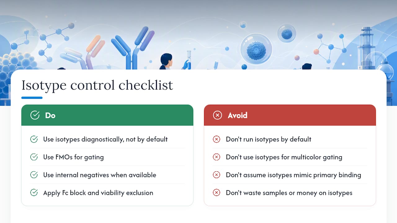
The labs that get this right treat isotype controls as a diagnostic reagent — something you reach for when something looks wrong, not something you run by default in every experiment. FMOs for gating, internal negatives where available, and rigorous Fc blocking and viability exclusion will give you cleaner, more reproducible data than a matched isotype tube ever will. Document your control strategy explicitly in your methods: which controls you used, why, and what each one revealed. Reviewers who understand flow cytometry will respect that more than a reflexive isotype lane.
Mayflowerbio supports your antibody and reagent needs
Getting isotype control selection right starts with having reagents you can trust lot to lot. Mayflowerbio carries a broad catalog of conjugated and unconjugated antibodies, including matched isotype controls, with lot-specific datasheets and QC documentation available on request. Whether you need a PE-conjugated mouse IgG1 isotype, an Fc blocking reagent, or a viability dye to complete your panel, the catalog is searchable by host species, Ig class, and conjugate.
 (Image content describing the Mayflowerbio antibody catalog and support services; no numerical data present)
For labs designing multicolor panels or moving into multiplex workflows, Mayflowerbio’s technical team can help you map your panel, identify the right controls, and confirm lot compatibility before you run. The reagent innovation resources on the site also cover assay design decisions that affect control selection upstream. Browse the antibody catalog or contact the technical support team to request lot documentation and datasheet access for your specific reagents.
Sources
- How to Identify Problems with Flow Cytometry Experiment Design – Bad Data Part 3 | Cytometry and Antibody Technology
- Pubmed
- Ncbi
FAQ
What is the point of an isotype control?
An isotype control reveals nonspecific binding and Fc receptor engagement by an antibody of the same class and conjugate as the primary, but with no relevant target in the sample. It does not set gates in multicolor panels and does not replicate the primary antibody’s nonspecific binding reliably across lots.
How do you choose an isotype control?
Match the primary antibody on four attributes: host species, immunoglobulin class and subclass, clonality (monoclonal or polyclonal), and conjugate or fluorochrome. Then titrate the isotype to the same working concentration as the primary before running the experiment.
What factors affect isotype control selection?
Host species, Ig class and subclass, clonality, conjugate, fluorochrome-to-protein ratio, and lot consistency all affect whether an isotype control accurately reflects the primary antibody’s background. Sample type, fixation, and Fc receptor expression on target cells also influence how much nonspecific signal the control will show.

What is an example of an isotype control?
A mouse IgG1 kappa PE-conjugated antibody used as an isotype control for a mouse IgG1 kappa PE-conjugated anti-CD4 primary antibody is a correctly matched example. Both share host species, subclass, clonality, and fluorochrome.
When should you use an FMO instead of an isotype control?
Use an FMO control whenever you are setting gates in a panel with three or more fluorochromes, especially for dim markers or markers with continuous expression. FMOs account for spillover and signal spreading from other channels; isotype controls do not, making them unreliable for gate placement in multicolor experiments.




