

Antibody Cross-Reactivity: Causes, Detection, and Fixes
Antibody cross-reactivity happens when an antibody binds an epitope other than its intended target, producing false bands, inflated signal, or misleading titers. It corrupts Western blots, ELISAs, IHC, and serology alike unless you control for it directly. The fastest fixes: run a no-primary control on every new lot, swap in a cross-adsorbed secondary or a directly conjugated primary when multiplexing, and confirm specificity in your exact assay before trusting the result.
TL;DR:
- Cross-reactivity occurs when antibodies bind structurally similar off-target epitopes, leading to false bands, misleading signals, or inflated titers across assays.
- Validation methods like peptide competition, knockout controls, and serial dilutions are essential to confirm antibody specificity within your experimental conditions.
- Choosing primary antibodies from different host species, using directly conjugated primaries, and selecting cross-adsorbed secondaries can significantly reduce cross-reactivity risks.
- Multiplex panels and serology are especially prone to cross-reactivity issues, which can cause incorrect diagnoses or misinterpretation of immune responses without proper orthogonal confirmation.
- Sourcing validated reagents and thoroughly documenting assay conditions before experimentation help prevent and troubleshoot cross-reactivity-related data problems.
Table of Contents
- What Causes Antibody Cross-Reactivity at the Molecular Level
- How Cross-Reactivity Shows Up in Western Blot, ELISA, IHC, and Serology
- How to Test for Antibody Cross-Reactivity in Your Assay
- Reagent and Design Choices That Reduce Cross-Reactivity Risk
- What Cross-Reactivity Means for Serology and Clinical Interpretation
- A Pre-Publication Validation Checklist for Antibody-Based Results
- Where Cross-Reactivity Problems Usually Come From, and Where to Get Help
- Reagents That Help You Control Cross-Reactivity Before It Costs You Data
- Sources
- FAQ
What Causes Antibody Cross-Reactivity at the Molecular Level
Cross-reactivity is not a manufacturing defect. It is a predictable consequence of how antibodies actually recognize antigen, and understanding the mechanism tells you which experiments will catch it.
An antibody’s paratope, the binding surface formed by its complementarity-determining regions, recognizes a three-dimensional shape rather than a unique molecule. When different proteins present structurally similar surfaces, the paratope can bind them too, reflecting that antibodies are specific to shapes which can recur across different proteins. The NIH’s overview of specificity and cross-reactivity makes this point directly: naming an antibody after its immunogen creates a false impression of exclusivity. In reality, a single antibody typically binds a spectrum of structurally related epitopes, each with different affinity.
Affinity and specificity are separate properties, and conflating them is one of the most common errors in antibody selection. A high-affinity antibody binds its intended target tightly but may still exhibit weaker detectable binding to related structures. Low-affinity, high-avidity interactions (multiple weak contacts adding up) can produce a signal just as strong as a single high-affinity interaction, which is part of why avidity matters as much as affinity when you’re troubleshooting an unexpected band.
Epitopes come in two structural flavors, and they behave differently under cross-reactivity testing. Linear epitopes are short, contiguous amino acid stretches, often 6 to 8 residues, that remain accessible even when a protein is denatured, which is why linear epitope antibodies tend to perform reliably in Western blot. Conformational epitopes depend on the folded, three-dimensional structure of the native protein, so an antibody raised against a folded antigen may fail entirely once the protein is denatured with SDS, or conversely may cross-react with an unrelated protein that happens to fold into a similar conformational patch. Peptide mimicry experiments using phage-displayed peptide libraries have mapped this directly: unrelated peptide sequences can adopt structures that mimic a genuine linear epitope closely enough to fool the paratope, a finding documented in structural cross-reactivity studies using systematic epitope mapping.

Then there’s polyspecificity, a property some antibodies have that goes beyond simple epitope overlap. A polyspecific antibody can adopt more than one paratope conformation, allowing a single antibody molecule to bind genuinely different, structurally unrelated antigens. The definitive review on this, covering specificity, polyspecificity, and heterospecificity in antibody-antigen recognition, notes that predicting which antibodies are polyspecific from sequence alone remains unreliable. Structural data, crystal structures or cryo-EM of the antibody bound to each ligand, is still the most trustworthy way to confirm which residues actually make contact. Interestingly, not everyone treats broad reactivity as a flaw: some newer array-based detection strategies deliberately exploit controlled cross-reactivity as a design feature rather than a liability, though for diagnostic and quantitative work, unplanned cross-reactivity remains a problem to eliminate, not a feature to embrace.
How Cross-Reactivity Shows Up in Western Blot, ELISA, IHC, and Serology
Cross-reactivity can be subtle and present in plausible-looking data, so assay-specific pattern recognition is crucial to detect it.
Western blot is usually the most forgiving assay for catching cross-reactivity, because molecular weight gives you an immediate sanity check. A band at the expected size is reassuring but not proof; a band at an unexpected size is a clue, not a verdict. Post-translational modifications shift apparent molecular weight, splice variants change it legitimately, and proteolytic degradation can produce a lower band that looks like cross-reactivity but isn’t. The practical lesson from antibody validation guidance for Western blot is that blocking reagent choice alone can change your result. Milk-based blockers may introduce background due to biotin and casein-derived peptides, while nonmammalian blocking agents can reduce background from mammalian-reactive antibodies in fluorescent multiplex blots.
ELISA and sandwich immunoassays manifest cross-reactivity differently, often through bridging rather than direct mis-binding. If capture and detection antibodies both recognize an off-target protein, or if a patient sample contains heterophile antibodies that physically bridge the two assay antibodies, you get a signal with no real analyte present. Watch for:
- Nonlinear dilution curves, where diluting the sample doesn’t proportionally reduce signal, a classic heterophile-interference signature
- Unexpectedly high signal in known-negative control samples
- Discordant results between two ELISA kits measuring the supposedly same analyte
IHC and IF carry a distinct cross-reactivity risk: secondary antibody and serum protein background. Endogenous immunoglobulins in tissue, Fc receptor binding, and cross-species serum reactivity (a secondary raised against one species reacting with endogenous IgG from another) all produce staining that has nothing to do with your primary antibody’s target.
Multiplex panels and serology compound every one of these issues. Panel-based serology, where a sample is tested against multiple related antigens simultaneously, often produces paradoxical results: a patient may show the highest titer against a serovar that isn’t actually the infecting one. A case study on the microscopic agglutination test for Leptospira demonstrated exactly this, finding that host species and lab-specific factors systematically alter cross-reactivity profiles, enough that the highest antibody titer in a panel cannot be assumed to identify the true infecting strain. Heterophile antibody interference compounds the problem in clinical sandwich assays for biomarkers like cardiac troponin, producing results that look clinically significant but aren’t.
How to Test for Antibody Cross-Reactivity in Your Assay
You cannot trust a datasheet’s specificity claim in your hands, in your buffer, on your instrument. Validation guidance is blunt about this: there is no guarantee an antibody validated elsewhere will be specific in your assay, because blocking reagent, sample matrix, and assay format all change antibody behavior. Here’s a prioritized sequence for confirming whether cross-reactivity is actually happening, and how much it’s affecting your data.
- Run peptide or antigen competition first. Pre-incubate the primary antibody with excess free antigen or the specific immunizing peptide before applying it to your sample. A true, specific signal should drop sharply, often 80% or more, while cross-reactive background persists because the competing peptide doesn’t bind the off-target site. No drop at all after competition is a red flag that your “specific” band might be an artifact entirely.
- Use a knockout, knockdown, or recombinant control. Nothing confirms target identity faster than a genetic negative. If a band or signal persists in a CRISPR knockout or siRNA knockdown line, it isn’t your target, full stop. Recombinant protein spiked into a blank matrix gives you the inverse confirmation: a clean positive control at the right molecular weight.
- Check serial dilution linearity. Dilute your sample across a 2 to 4 fold series and plot signal against dilution. A genuine, specific interaction produces a linear or near-linear relationship on a log scale. Heterophile interference and nonspecific bridging often produce flat or erratic curves that don’t respond proportionally to dilution.
- Switch platforms for orthogonal confirmation. If ELISA gives a suspicious result, confirm with a different detection chemistry, chemiluminescence versus colorimetric, or an entirely different method like mass spectrometry or PCR-based detection for the same target. Agreement across orthogonal platforms is much stronger evidence than a clean-looking result on a single platform.
- Run the mismatched-secondary and pre-adsorption test. Apply your secondary antibody alone, with no primary, to check for direct tissue or membrane binding. Separately, pre-adsorb the secondary against serum or IgG from the “wrong” species before use; a properly cross-adsorbed secondary should show minimal residual reactivity against non-target species immunoglobulin.
Quantify what you find rather than eyeballing it. Percent signal reduction after competition (specific signal should fall by the majority of its intensity), a competition curve with a defined IC50-style inflection point, and a residual cross-reactivity percentage against off-target species are all metrics you can put in a validation report. As a rough decision rule: if competition or knockout controls reduce signal by less than half, treat the antibody as unacceptable for quantitative work until you resolve why.
Pro Tip: If a phospho-specific antibody shows a band you can’t explain, treat the sample with lambda phosphatase before running it again. If the band disappears, you’ve confirmed the antibody genuinely requires that modification rather than cross-reacting with the unmodified protein or an unrelated phosphoprotein.
Reagent and Design Choices That Reduce Cross-Reactivity Risk
Most cross-reactivity problems get solved before the experiment starts, not after the data comes back strange. Reagent selection and workflow design carry more weight than any downstream fix.
- Choose primary antibodies from distinct host species for multiplex work. If you’re staining for three targets simultaneously, primaries raised in rabbit, mouse, and goat (rather than two from the same species) let you use species-specific secondaries without ambiguity about which secondary is binding which primary.
- Use directly conjugated primaries when multiplexing gets complicated. Skipping the secondary antibody step entirely removes an entire category of cross-reactivity risk, at the cost of typically weaker signal amplification.
- Select cross-adsorbed secondaries whenever your sample contains immunoglobulins from more than one species. Cross-adsorption is a purification process where the secondary antibody is run against immobilized IgG from other species to strip out any antibody population that would react with them. Manufacturer testing confirms cross-adsorbed secondaries substantially reduce off-target species reactivity compared to non-adsorbed versions, which matters most in tissue with high endogenous immunoglobulin content, like spleen, lymph node, or inflamed samples.
- Pick your blocking reagent deliberately, not by habit. Nonfat dry milk is cheap and effective for most total-protein blots but contains biotin and phosphoproteins that interfere badly with phospho-specific antibodies and biotin-streptavidin detection systems. BSA is often the safer default for phospho-work. Nonmammalian blocking agents can reduce background specifically from mammalian-reactive antibodies in fluorescent multiplex applications.
- Avoid simultaneous antibody cocktails when you can sequence instead. Experienced multiplex labs increasingly favor sequential primary incubation with thorough washes between steps over one-shot cocktails, precisely because unpredictable antibody-antibody or antibody-Fc receptor interactions in a mixed cocktail are hard to troubleshoot after the fact.
- Know when to abandon an antibody rather than patch around it. If a polyclonal keeps producing inconsistent cross-reactivity across lots, switching to a monoclonal targeting a well-mapped epitope, or requesting the supplier’s actual validation data (knockout blots, competition data, not just a datasheet claim), is usually faster than continuing to troubleshoot.
Pro Tip: Keep a running log of which blocking reagent and secondary lot you used for every antibody that’s given you trouble. Cross-reactivity problems that look random from run to run often turn out to correlate with a specific reagent lot once you have six months of notes to compare.
What Cross-Reactivity Means for Serology and Clinical Interpretation
Serology interpretation is where cross-reactivity stops being a bench annoyance and starts affecting patient management. The core trap: assuming the antigen with the highest titer in a multi-antigen panel identifies the true cause.
The Leptospira microscopic agglutination test case study is the clearest illustration available: researchers found that host species and even which laboratory ran the assay changed which serovar produced the dominant titer, independent of which strain actually caused infection. Titers from one lab are not automatically comparable to titers from another, and a “positive” result against one antigen in a panel doesn’t rule out cross-reactive binding driving that signal.
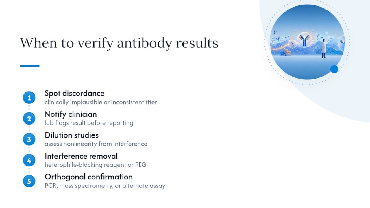
Heterophile antibodies deserve specific attention because they cause a particular, dangerous pattern: a result that looks clinically real, is internally consistent, and simply won’t budge on repeat testing. This is a legitimate clinical red flag, not an assay quirk. Heterophile interference in cardiac biomarker testing has caused documented cases of unnecessary intervention when troponin results didn’t match the clinical picture, and the recommended verification path includes dilution studies, heterophile-blocking reagent treatment, PEG precipitation, and confirmatory testing on an alternative platform.
The practical rule for any discordant or clinically implausible antibody-based result: confirm with an orthogonal, ideally molecular method (PCR for infectious targets, mass spectrometry for protein identity) before treating the immunoassay result as definitive. Lab staff flagging a suspicious result to the ordering clinician, rather than reporting a number with no context, is the difference between catching heterophile interference early and having it drive a wrong clinical decision.
A Pre-Publication Validation Checklist for Antibody-Based Results
Before you trust an antibody-based result enough to put it in a manuscript or a clinical report, run it through a fixed sequence rather than an ad hoc gut check.
- Run the core control panel for every new lot and every new assay format: no-primary control, isotype control matched to your primary’s species and class, single-plex comparison if you’re running multiplex, and a mismatched-secondary test.
- Confirm with at least one orthogonal method: recombinant protein spike-in, a knockout or knockdown line if available, peptide competition, or a different detection platform entirely.
- Document everything that could explain a discrepancy later: antibody lot number, blocking reagent and lot, exposure settings or scan parameters, full dilution series data, and raw images, not just cropped bands.
- Set a decision threshold before you look at the data, not after. A common working standard is that specific signal should drop by more than half under competition or disappear in a genetine knockout; residual cross-reactive signal above that threshold means the reagent needs replacement or a change in dilution, blocking, or detection chemistry, not a footnote explaining it away.
A useful benchmark: the antibody validation literature is consistent that specificity is not portable between labs or even between assay formats within the same lab. A Western blot validation review makes the case plainly: the burden of proof sits with the user running the assay, not the datasheet the antibody shipped with.
Where Cross-Reactivity Problems Usually Come From, and Where to Get Help
The pitfalls we hear about most often are predictable: a phospho-antibody that suddenly stops working after a switch to a new milk lot, a multiplex IHC panel with bleed-through nobody caught until publication review, or a secondary antibody that wasn’t cross-adsorbed showing up as unexplained background in mixed-species tissue.
Every one of those is fixable with the controls covered above, and it’s worth running them before you commit bench time to a full experiment. Product-specific validation notes, including which applications and species an antibody has actually been tested against, are worth checking before you order rather than after a failed blot. If your controls point to a genuine specificity problem rather than a technique issue, our technical support team can help you work through it.
— Alina
Reagents That Help You Control Cross-Reactivity Before It Costs You Data
Choosing the right antibody format up front avoids most of the cross-reactivity headaches described above, and it’s a lot cheaper than repeating a failed experiment.

Mayflowerbio’s antibody catalog includes application-specific validation notes so you can check host species, tested applications, and cross-reactivity data before you order rather than after a failed blot. For labs running multiplex panels, our AimPlex multiplex assay platform is built around bead-based detection designed to minimize the antibody bridging and cross-talk that plague crowded multiplex ELISA formats, and our guide on multiplex assay design principles walks through panel configuration choices that reduce interference risk before you run your first plate. None of this replaces validating a reagent in your own hands with the controls covered in this article, that responsibility stays with your lab, but starting from a well-documented antibody saves you a round of troubleshooting. Browse the antibody catalog or reach out to technical support to talk through which format fits your panel.
This article is general information, not a substitute for advice from a qualified doctor. Consult a qualified healthcare professional about your own circumstances before acting on anything here.
Sources
- Antibody validation for Western blot: By the user, for the user
- Navigating cross-reactivity and host species effects in a serological assay: A case study of the microscopic agglutination test for Leptospira serology
- Specificity, polyspecificity, and heterospecificity of antibody-antigen recognition
FAQ
What does it mean when an antibody is cross-reactive?
A cross-reactive antibody binds an epitope on a protein other than its intended target, usually because that off-target epitope shares structural similarity with the true antigen. It can produce false-positive bands, background staining, or misleading titers depending on the assay.
What does cross-reactivity mean in immunology generally?
Cross-reactivity describes any immune molecule, antibody or T-cell receptor, recognizing more than one distinct antigen. For antibodies, it stems from epitope-paratope complementarity being based on shape rather than a single unique sequence, so structurally related molecules can trigger the same binding event.
What does it mean if an antibody is reactive?
“Reactive” means the antibody produced a detectable signal against a given antigen in an assay. Reactive is not the same as specific: an antibody can be reactive against your true target and also reactive against an unrelated protein if the two share a compatible epitope structure.
Are there clinical symptoms linked to antibody cross-reactivity?
Cross-reactivity itself causes no symptoms since it’s an assay or laboratory phenomenon, not a patient condition. Its real-world impact is diagnostic: a cross-reactive result can trigger an incorrect diagnosis, an unnecessary confirmatory workup, or a delay in identifying the correct condition when a heterophile or cross-reactive signal is mistaken for a true positive.
How do I know if my antibody’s cross-reactivity is a real problem?
Run peptide competition or a knockout control; if the signal in question doesn’t drop substantially, it’s likely cross-reactive rather than specific. Pair that with a mismatched-secondary test to rule out secondary-antibody contributions before concluding the primary itself is the source.




