

4 Strategies for Growth Factor Carriers That Cut Burst Release in Labs
Growth factor carriers exist because free proteins fail fast: they diffuse away, degrade within minutes, and demand supraphysiological doses to get anywhere near a therapeutic effect. The strategies that actually work fall into four buckets: affinity sequestration that mimics how the extracellular matrix naturally holds growth factors, physical encapsulation in particles or hydrogels, covalent immobilization onto a scaffold, and engineered growth factors carrying built-in matrix-binding domains. Each solves the same problem differently: keeping the signal local, active, and timed to biology instead of dumped in a single bolus.
TL;DR:
- Growth factor delivery systems must balance local retention, controlled release, and bioactivity preservation to replicate natural healing signals effectively.
- Hydrogels, nanoparticles, and natural matrices are the most common materials, but each offers different trade-offs in degradation control, loading capacity, and biocompatibility.
- Combining encapsulation with affinity-based binding significantly reduces burst release and improves retention at the target site.
- Optimizing release kinetics to match tissue healing timelines is more critical than simply maximizing loading capacity.
- Early validation of bioactivity retention and careful consideration of sterilization and scale-up are essential to avoid costly setbacks in development.
Table of Contents
- Why Growth Factor Carriers Are Necessary
- Material Classes Used as Growth Factor Carriers
- How Do You Control Growth Factor Release and Presentation?
- Design and Evaluation Criteria for Carrier Systems
- Applications Where Carriers Make the Difference
- Engineering Advances in Growth Factor Carrier Design
- What I’d Prioritize If I Were Starting This Work Today
- How Mayflower Bioscience Supports Growth Factor Carrier Research
- Key Reviews and Primary Sources to Read Next
- Sources
- FAQ
Why Growth Factor Carriers Are Necessary
Most growth factors were never built to survive outside a tightly regulated tissue microenvironment. VEGF, BMPs, and TGF-β normally act over microns, for hours, at concentrations the body fine-tunes moment to moment. Inject them as a free bolus and you get the opposite: rapid dilution, proteolytic breakdown, and a spike-then-crash exposure curve that bears no resemblance to native signaling.
Direct bolus injections are inefficient largely because of rapid diffusion and enzymatic degradation, which is why clinical protocols using free growth factor often require supraphysiological doses that raise side-effect risk. BMP-2 in spinal fusion is the textbook cautionary example: effective at high concentrations, but linked to ectopic bone formation and inflammation when carrier control was inadequate.
Carriers change the pharmacokinetics entirely. They:
- Shield the protein from serum proteases and extend its functional half-life at the delivery site
- Keep concentration localized instead of letting the factor diffuse into systemic circulation
- Allow dose reduction by matching release rate to the tissue’s actual consumption rate
- Convert a single spike exposure into a sustained or staged release profile that tracks the healing timeline
Controlled release systems matter because they align delivery with how the body naturally produces growth factors during repair, which is exactly what makes VEGF, BMP, and TGF-β delivery so dependent on getting the carrier right, not just the dose.
Material Classes Used as Growth Factor Carriers
No single material wins across every application. The choice depends on the target tissue, the release window you need, and how much you’re willing to trade loading capacity for degradation control.
Hydrogels remain the default for soft tissue and bone applications because mesh size and crosslink chemistry are both tunable. Protease-sensitive crosslinks let the hydrogel degrade in step with cellular infiltration, and affinity motifs (often heparin-mimetic sequences) can be grafted in to slow diffusion without chemically altering the growth factor itself.
Synthetic polymer micro and nanoparticles, particularly PLGA and PEG-based systems, offer reliable, tunable degradation kinetics through polymer molecular weight and copolymer ratio. Their main liability is burst release: a poorly formulated particle can dump 40 to 60 percent of its payload in the first hours, which defeats the purpose of encapsulation.
Nanoparticles and mesoporous silica bring high surface area and highly tunable pore chemistry, which supports dense peptide and growth factor loading. Silica-based carriers have shown improved osteogenic differentiation when BMP-2 or bioactive peptides are immobilized on the particle surface, largely because the porous structure buffers release rate against simple diffusion.
Lipid-based carriers, including liposomes and nanostructured lipid carriers, protect against protein denaturation and accommodate amphiphilic loading strategies that aqueous systems can’t handle as cleanly.
Natural ECM-derived matrices — fibrin, gelatin, collagen — win on biocompatibility. Processing conditions are mild enough to preserve native protein conformation, and cells recognize these matrices through the same integrin pathways they’d use in vivo.
Pro Tip: Pre-encapsulate the growth factor in a nanoparticle before embedding that particle in a hydrogel. Combining encapsulation with affinity-based matrix binding gives you both a diffusion barrier and a retention mechanism, which cuts burst release far more than either strategy alone.
How Do You Control Growth Factor Release and Presentation?
Choosing a release mechanism is really choosing a trade-off between simplicity and control. Five mechanisms dominate the literature, and most translational systems combine at least two.
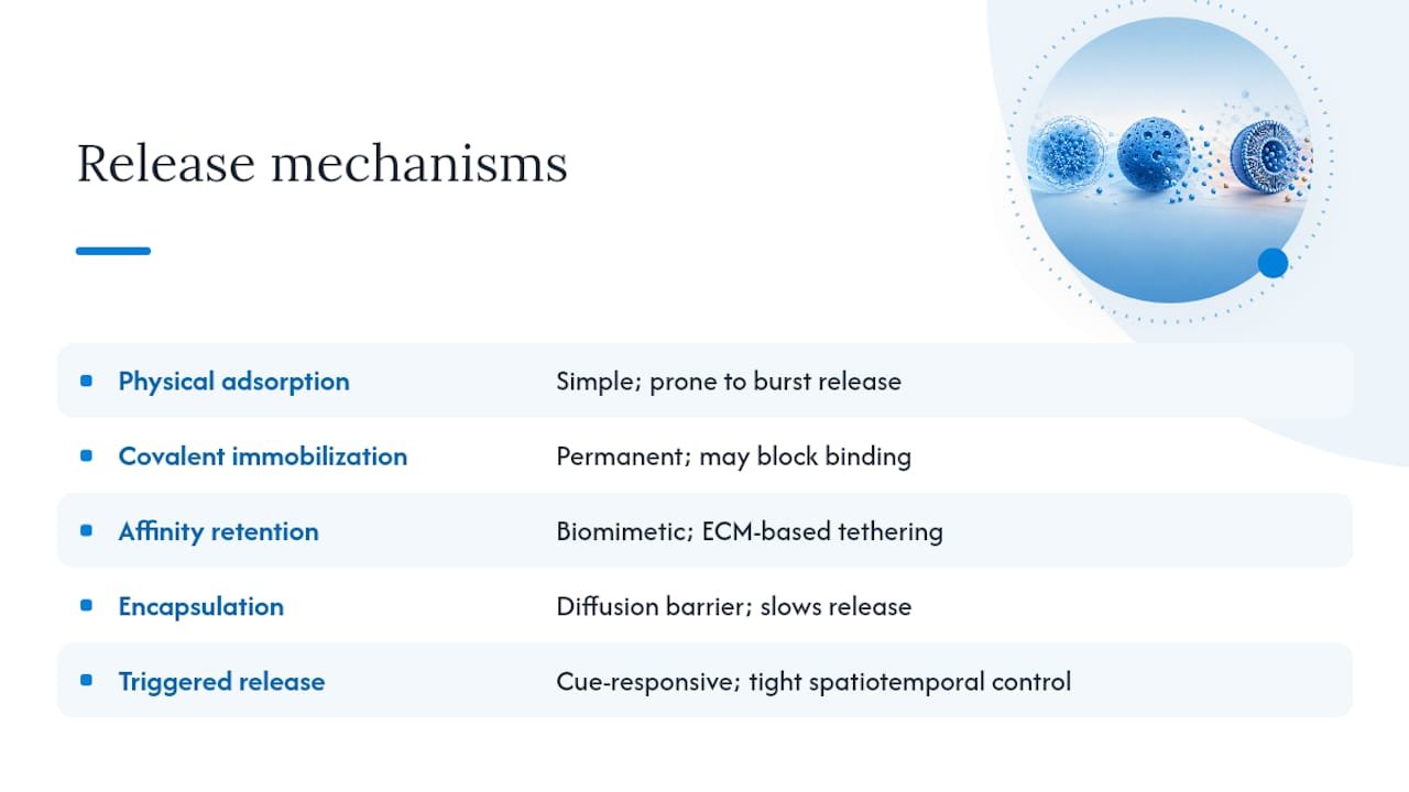
Physical adsorption is the simplest approach: soak the scaffold in growth factor solution and let electrostatic or hydrophobic interactions hold it in place. It’s fast and cheap, but adsorbed protein is loosely bound and prone to burst release within the first day. Combining adsorption with a secondary encapsulation step is a common fix.
Covalent immobilization tethers the growth factor permanently to the material, which gives durable, long-term presentation. The trade-off is real: covalent bonding can block the receptor-binding epitope or restrict the conformational flexibility a receptor needs to trigger signaling, so tether-site selection matters as much as the chemistry itself.
Affinity-based retention using heparin, glycosaminoglycan mimetics, or ECM-binding peptides recreates how the native matrix sequesters growth factors in vivo. This is arguably the most biomimetic mechanism available and underlies much of the current push toward engineered growth factor fusions.
Encapsulation in particles or core-shell architectures adds a diffusion barrier that slows release independent of binding chemistry, which is why layering encapsulation on top of affinity binding tends to outperform either method alone.
Triggered release systems respond to enzyme activity, pH shifts, light, or magnetic fields, releasing payload on demand rather than on a fixed timetable. These systems are still mostly preclinical but offer the tightest spatiotemporal control of any category, matching release to a biological cue rather than a preset degradation rate.
Design and Evaluation Criteria for Carrier Systems
Comparing carrier systems requires more than “does it release the growth factor.” A practical evaluation framework, echoed across the field, breaks the problem into biological considerations, material selection, and delivery strategy as three linked design axes rather than independent choices.
Four metric categories separate a rigorous carrier study from a superficial one:
- Release kinetics: encapsulation efficiency, burst fraction in the first 24 to 48 hours, cumulative mass released over the study window, and functional half-life in physiologically relevant media rather than plain buffer.
- Bioactivity retention: receptor phosphorylation assays, proliferation or migration readouts in relevant cell types, and functional endpoints that confirm the released factor still signals correctly, not just that it’s present.
- Safety and compatibility: cytotoxicity of the carrier and its degradation products, immunogenicity risk, and confirmation that sterilization method hasn’t altered either the matrix or the growth factor’s conformation.
- Manufacturing readiness: batch-to-batch reproducibility, scalability of the fabrication process, and compatibility with regulatory-grade quality control.
Reviews increasingly frame spatiotemporal delivery, meaning sequential and localized release timed to tissue biology, as the variable that separates carriers that work in a dish from carriers that work in an animal. A carrier that scores well on loading efficiency but poorly on bioactivity retention isn’t actually a better system; it’s a worse one with better marketing numbers.
Applications Where Carriers Make the Difference
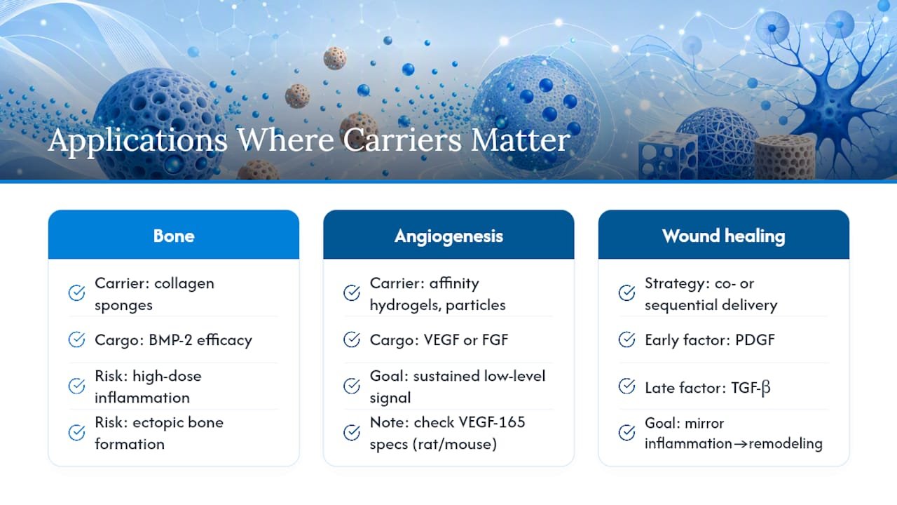
Bone regeneration is where carrier-dependent growth factor therapy has the longest clinical track record. BMP-2 delivered on collagen sponges achieves real efficacy, but clinical experience also exposed the downside: without tighter carrier control, effective doses ran high enough to trigger inflammatory and ectopic bone complications, which is exactly the dosing risk carriers are meant to eliminate.
Angiogenesis work leans heavily on affinity-functionalized hydrogels and sustained-release particles carrying VEGF or FGF, since new vessel formation depends on a sustained low-level signal rather than a spike. You can review VEGF-165 recombinant protein specifications for rat and mouse models when designing these systems.
Wound healing studies increasingly favor co-delivery or sequential release, pairing an early-phase factor like PDGF with a later-phase factor like TGF-β to mirror the natural inflammatory-to-remodeling transition rather than releasing everything at once.
The recurring translational snag across all three applications: animal models routinely respond to growth factor doses that don’t scale linearly to human tissue volume, and carrier systems validated in small-animal defects frequently need substantial reformulation before larger preclinical or clinical studies.
Engineering Advances in Growth Factor Carrier Design
The most interesting work right now isn’t in the carrier material at all. It’s in redesigning the growth factor itself. Fusing ECM-binding domains, often derived from placental growth factor or fibronectin fragments, directly onto a growth factor increases local retention and has repeatedly enabled therapeutic efficacy at lower doses in preclinical bone and wound models.
Three approaches define this frontier:
- Functionalizing carrier surfaces with heparin-mimetic or fibronectin-derived peptides to recreate native sequestration without modifying the growth factor
- Fusing matrix-binding domains directly onto the recombinant protein, an accessible route for labs already running standard cloning and expression workflows
- Building “smart” carriers that actively resist proteolytic degradation and time their release to match the biological phase of repair, rather than relying on passive diffusion
Pro Tip: If you’re screening ECM-binding fusion constructs, validate bioactivity retention with a receptor phosphorylation assay before moving to any in vivo model. A fusion that binds matrix well but loses receptor affinity is a common and costly failure mode. Mayflower Bioscience supplies recombinant growth factors and assay reagents suited to exactly this kind of bench-stage validation.
What I’d Prioritize If I Were Starting This Work Today
Run bioactivity retention assays before you touch an animal model. A carrier that looks elegant in a release-kinetics plot is worthless if the protein loses receptor affinity along the way, and that failure mode is cheap to catch early and expensive to catch late.
Match your release kinetics to the tissue’s actual healing timeline, not to whatever loading capacity your material can technically hit. Maximal loading is a vanity metric if the release profile doesn’t track biology.
And plan for sterilization and scale-up from day one. Reformulating a carrier after it’s already validated in vivo, because your sterilization method degraded the matrix, is one of the most avoidable setbacks in this field.
— Alina
How Mayflower Bioscience Supports Growth Factor Carrier Research
Mayflowerbio is the practical starting point for labs building and validating carrier systems, not another materials vendor asking you to guess at specifications. We supply recombinant growth factors, including VEGF-165, TGF-α, and FGF-21, alongside the assay reagents and antibodies needed to confirm bioactivity survived your encapsulation or immobilization step.

Whether you’re validating burst release with a phospho-specific antibody panel or need bulk recombinant protein for a scaffold screening study, our cell biology reagent catalog covers the bench-stage tools most carrier projects actually run through. Technical specifications, certificates of analysis, and bulk or custom quantities are available on request. Visit the product catalog to check specifications or contact our technical support team for a quote on your next carrier validation study.
Key Reviews and Primary Sources to Read Next
For deeper reading, start with the PMC review on controlled delivery strategies for a broad mechanistic overview, the engineering-focused PMC review for matrix-binding fusion design, and the ScienceDirect design-platform paper for a structured evaluation framework.
This article is general information, not a substitute for advice from a qualified doctor. Consult a qualified healthcare professional about your own circumstances before acting on anything here.
Sources
- Novel biomaterial strategies for controlled growth factor delivery
- Engineering growth factors for regenerative medicine applications
FAQ
Does growth factor therapy actually work?
Yes, but efficacy depends heavily on delivery. Reviews consistently link successful outcomes to the presence of a carrier system that controls spatiotemporal presentation rather than free protein delivered alone.
What are the three main types of growth factors used in carrier research?
VEGF, BMPs, and TGF-β are the three most extensively studied in carrier literature, covering angiogenesis, bone regeneration, and tissue remodeling respectively. FGF and PDGF are also widely used, particularly in wound healing and vascularization work.
Are growth factors or exosomes the better delivery approach?
They solve different problems: growth factor carriers deliver a defined, known signaling molecule with controllable kinetics, while exosomes deliver a complex, less-defined cargo mixture. Neither is universally “better;” the choice depends on whether your application needs a single characterized signal or a broader paracrine effect.
What makes a growth factor carrier design effective?
Effectiveness comes down to matching release kinetics to the tissue’s healing timeline while preserving bioactivity, not maximizing loading capacity. Carriers that combine encapsulation with affinity-based retention consistently outperform single-mechanism systems on burst control.




