
300–2,000×g Spinoculation Protocol for Biomedical Researchers
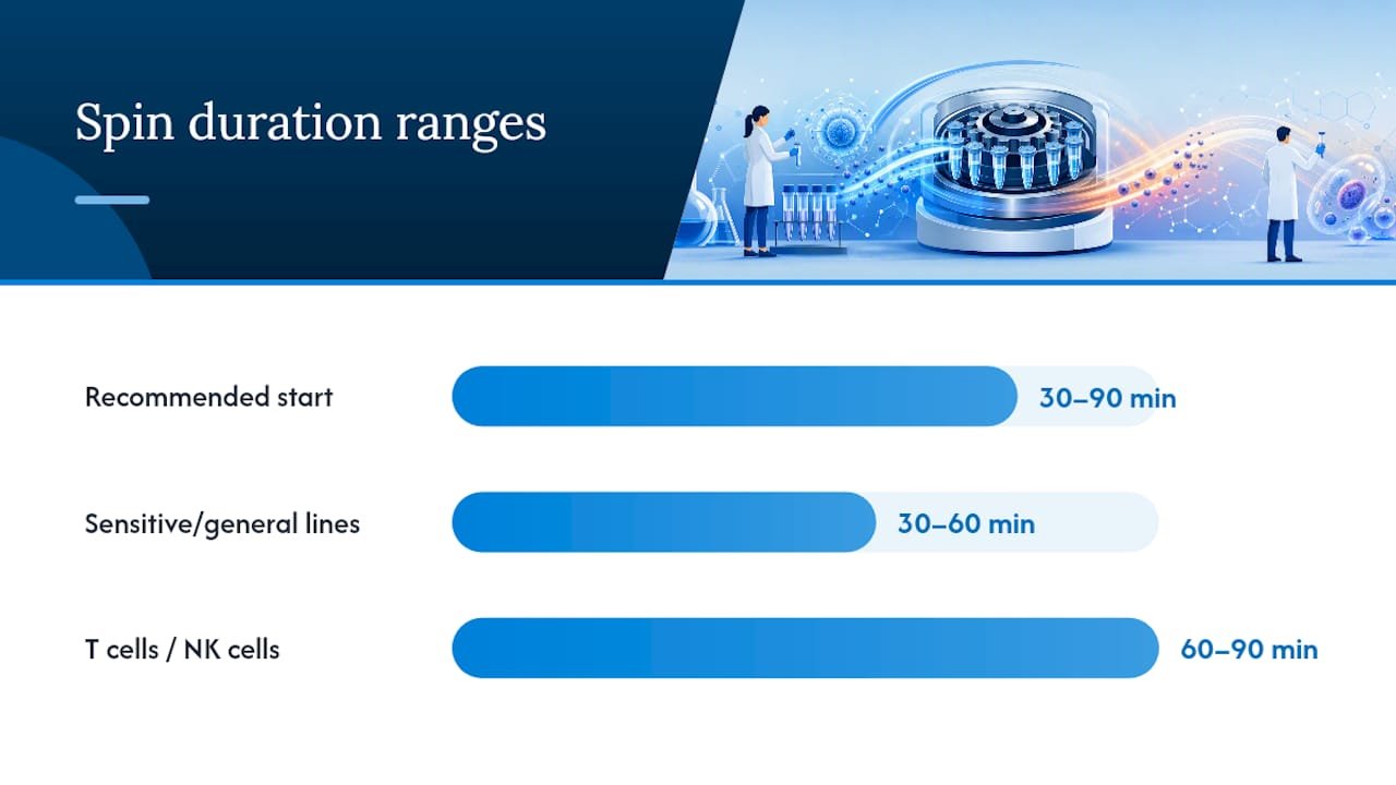
Start at 1,000 to 2,000×g for 60 to 90 minutes at room temperature or 32°C, with 4 to 8 µg/mL polybrene or a retronectin coat and expect a notable increase in transduction depending on your cell type. The tradeoff is real: higher g forces and longer spins stress cells and slow recovery, and the method doesn’t scale cleanly to bioreactor volumes. If your first run underperforms, check the troubleshooting and alternatives sections below before you assume the cells are simply resistant.
TL;DR:
- Retronectin-coated plates outperform polybrene alone, providing higher transduction rates and less cytotoxicity for sensitive primary cells.
- Proper viral titer validation through functional assays is critical to avoid under- or overdosing, which can reduce efficiency or increase toxicity.
- Temperature management involves balancing viral stability and cell metabolism by choosing room temperature or 32°C during spin, especially with longer durations.
- Careful control of biosafety procedures and equipment setup minimizes risk and ensures reliable results in spinoculation protocols.
Table of Contents
- What Do You Need Before Running a Spinoculation Protocol?
- How Do You Run a Spinoculation Protocol Step by Step?
- What Are the Optimal G-Force, Time, and Temperature Ranges?
- What’s the Best Way to Concentrate and Titer Virus for Spinoculation?
- Why Is My Transduction Efficiency Low or Toxicity High?
- What Biosafety Precautions Does Spinoculation Require?
- When Should You Use Static Transduction or Retronectin Instead?
- What I’d Actually Recommend Running First
- Where to Read the Original Protocols
- Sources
- FAQ
What Do You Need Before Running a Spinoculation Protocol?
Getting the materials right before you touch a pipette saves you from the most common failure mode in this technique: discovering mid-protocol that your rotor can’t hit the target g force, or that you diluted your virus past the point of a usable multiplicity of infection (MOI). Here’s what belongs on your bench.
Viral stock and enhancers. You’ll need concentrated lentivirus with a known functional titer, plus your chosen transduction enhancer. Polybrene remains the default at 4 to 8 µg/mL, but it comes with a cytotoxicity cost, especially in primary lymphocytes. Retronectin, coated onto plates or tubes at roughly 5 to 20 µg/cm², works through a different mechanism, colocalizing virus and cells on a fibronectin fragment rather than neutralizing surface charge. Data from a study on MUC1 CAR T-cell transduction found spinoculation plus retronectin hit 63.19% transduction efficiency, compared to 34.6% for spinoculation plus polybrene and just 10.23% for polybrene alone, with polybrene groups also showing higher cytotoxicity. If you’re working with sensitive primary cells, that’s the single most important materials decision you’ll make. Poloxamer-based and LentiBOOST-style chemical additives are a newer option; a quality-by-design study on CAR T manufacturing found these additives can increase transduction significantly on their own, and combined with optimized physical parameters, push overall gains substantially without hurting cell growth.
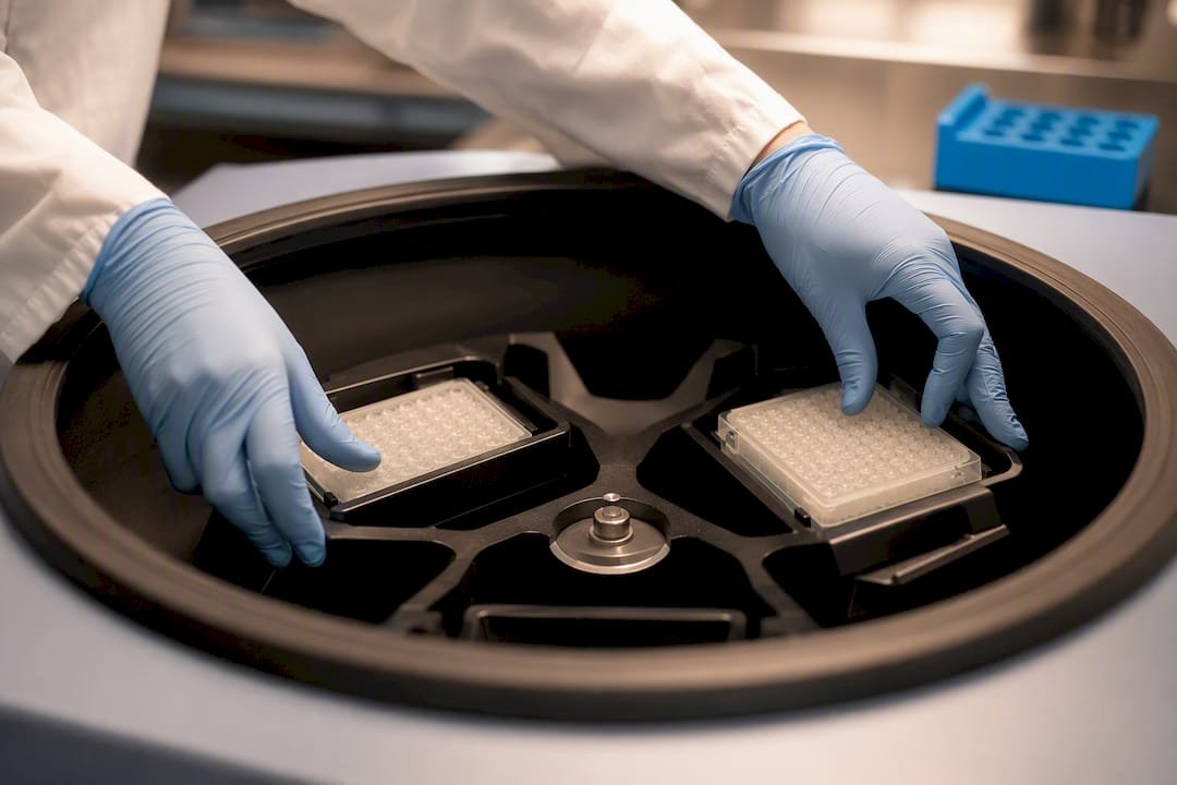
Labware. Twenty-four-deep-well pyramidal-bottom plates are the standard for small-scale plate spins because the pyramidal geometry concentrates cells and virus at a single low point rather than spreading them across a flat well bottom. Standard 24-well and 6-well plates work fine too, just expect slightly less local concentration. Whatever format you choose, balance your plates or tubes symmetrically across the rotor. Swinging-bucket rotors are strongly preferred over fixed-angle rotors for spinoculation. A fixed-angle rotor pellets cells against the side wall at an angle, which distorts the monolayer of cells the virus needs to contact evenly.
Equipment. You need a centrifuge that can hold your target g force at a controlled temperature, ideally with a plate-carrier rotor rated for the speeds you’re planning. A class II biosafety cabinet is required for all liquid handling involving lentiviral vectors, and you’ll want a standard 37°C, 5% CO2 incubator for recovery.
Controls and assays. Every spinoculation run needs a positive control, typically a GFP or luciferase-expressing lentivirus at a known titer, run in parallel with your experimental virus. Include a mock-transduced (no virus) well to set your flow cytometry gates and a viability dye, such as trypan blue or a fixable live/dead stain, to separate transduction failure from cell death.
- Concentrated lentivirus with a validated functional titer
- Polybrene (4 to 8 µg/mL) or retronectin-coated plates (5 to 20 µg/cm²)
- Chemical enhancer such as a LentiBOOST-type additive, if available
- 24-well deep-well pyramidal plates or standard 24-well/6-well plates
- Swinging-bucket centrifuge rotor rated for your target g force
- Biosafety cabinet and CO2 incubator
- GFP or luciferase reporter virus as a positive control
- Viability dye and mock-transduction control well
If you need reagents or transduction-grade viral stock to start building this kit, Mayflowerbio’s viral transduction and bioprocessing tools page covers the enhancer and reagent categories most protocols call for.
How Do You Run a Spinoculation Protocol Step by Step?
The core protocol below is built for 24-well and 6-well plate formats, with notes on adapting it for suspension bioreactor cultures. Follow it in order. Skipping the mock control or rushing the post-spin resuspension step is where most first attempts go wrong.
Cell preparation
- Count your cells and check viability with trypan blue or an automated counter. Anything under 90% viability going into the spin is a red flag; fix that before you add virus.
- If you’re working with T cells or NK cells, activate them first. T cells typically need 24 to 48 hours of anti-CD3/CD28 stimulation before transduction; unactivated T cells transduce poorly regardless of spin conditions because lentivirus requires actively dividing or metabolically active target cells for efficient integration.
- Plate cells at a density appropriate to your format. For 24-well plates, 0.5 to 1×10^6 cells per well is typical; for 6-well plates, scale up to roughly 1.5 to 2.5×10^6 cells per well. Suspension formats can run slightly denser since you’re not relying on adherent surface area.
Virus preparation
- Thaw your concentrated viral stock on ice, never at room temperature, and avoid refreezing any unused volume.
- Dilute the virus into your final transduction volume based on your target MOI (see the parameters section below for how to calculate this from your titer). Keep the total volume as small as your plate format allows. Diluting too aggressively lowers the local viral concentration cells actually experience during the spin, which undercuts your effective MOI even if the total particle count on paper looks adequate.
- Set up a dilution series alongside your main condition, typically MOI 1, 5, 10, and 20, plus a virus-free mock well. This lets you find your optimal MOI in the same run instead of guessing and repeating the whole experiment later.
- Add your enhancer now. If using polybrene, add it directly to the diluted virus at your chosen concentration (4 to 8 µg/mL final). If using retronectin, your plates should already be pre-coated and blocked before this step.
The spin
- Add the cell suspension to each well containing diluted virus, mixing gently by pipetting up and down twice. Don’t vortex.
- Seal plates with a breathable membrane or use plate-specific lids designed for centrifugation to prevent cross-contamination between wells.
- Balance the rotor carefully, opposing plates or tubes of equal weight. An unbalanced swinging-bucket rotor at 2,000×g is a real safety hazard, not just a data quality issue.
- Spin at your chosen g force and duration (see the parameters table in the next section for cell-type-specific windows). Room temperature works for most robust lines; primary lymphocytes and other sensitive cells often do better at 32°C, which balances viral stability against the metabolic slowdown of a full 4°C cold spin.
Post-spin handling
- After the spin, most protocols leave the viral supernatant on the cells rather than removing it immediately, particularly for suspension formats where aspiration is impractical. If you are working in an adherent or plate format and cytotoxicity is a concern, you can carefully remove roughly half the supernatant and replace it with fresh medium 4 to 6 hours post-spin.
- Return plates to the incubator at 37°C, 5% CO2, undisturbed for at least the first 24 hours. Resist the urge to check on them under the microscope during that window; the cell pellet formed by the spin needs time to redisperse naturally.
- At 24 hours, gently resuspend (for suspension formats) or do a half-media change (for adherent formats) to dilute out residual polybrene, which continues to exert some toxicity the longer it sits on cells.
Readout and assays
- Sample at 48 to 72 hours post-transduction for reporter readout. This window is standard because lentiviral integration and reporter gene expression need time to ramp up, but going much past 96 hours risks confounding your signal with cell division diluting episomal, non-integrated viral DNA.
- Run flow cytometry for GFP or your reporter of choice, gating against your mock-transduced control. For luciferase reporters, run a luminescence assay per your kit’s instructions.
- Check viability at the same timepoint using your dye of choice. A transduction efficiency number without a paired viability number tells you only half the story, since a spin that kills 40% of your cells while transducing 80% of survivors isn’t necessarily a win over a gentler protocol at 60% transduction and 90% viability.
Pro Tip: Run your MOI dilution series and your g-force optimization in the same 24-well plate the first time. You’ll burn one extra plate of cells, but you’ll walk away with a two-dimensional map of what actually drives your transduction efficiency instead of guessing which variable to adjust next.
For suspension bioreactor formats, the core logic holds, but a few things change. You typically can’t spin the entire bioreactor volume, so cells are pelleted in batches, exposed to concentrated virus and enhancer in a reduced volume for the spin step, then returned to the larger culture volume. Keep the spin-phase volume as concentrated as your rotor and tube format allow, since this is where you get the biggest gain over static transduction in suspension-adapted lines. Track viability more closely in these formats. The transfer between spin tubes and the bioreactor introduces extra shear and handling stress that plate-based protocols don’t have.
What Are the Optimal G-Force, Time, and Temperature Ranges?
The physical parameters you choose have more effect on outcome than any single reagent decision, and the right window depends heavily on how sensitive your cell type is.
For robust, adherent-adjacent lines and many suspension cell lines, 300 to 800×g for 30 to 90 minutes at room temperature is a reasonable starting window. For T cells and NK cells, published data supports going considerably higher. Research on primary immune cell transduction found that spinning at higher g forces and longer durations substantially increased transduction compared to no spin, without necessarily reducing T-cell growth. That’s a more aggressive window than many protocols default to, and it’s worth testing if your low-g runs are underperforming.
| Parameter | Sensitive/general suspension lines | T cells / NK cells |
|---|---|---|
| G-force | 300 to 800×g | 1,000 to 2,000×g |
| Duration | 30 to 60 minutes | 60 to 90 minutes |
| Temperature | Room temperature | Room temperature to 32°C |
| Typical enhancer | Polybrene 4 to 8 µg/mL | Retronectin or polybrene |
Temperature choice interacts with duration. A room-temperature spin held for 90 minutes exposes both cells and virus to more time at a temperature where lentiviral particles start losing infectivity. Pre-chilling to 4°C protects the virus but slows the metabolic activity that helps cells take up the vector, which is why many primary cell protocols split the difference at 32°C. If your titer is limited and you can’t afford loss of infectivity over a long spin, shortening the duration and raising the g force is usually a better tradeoff than lowering the temperature.
MOI planning deserves its own mention here because it’s the parameter most people get wrong before they ever reach the centrifuge. Functional titer (measured by flow cytometry on a reporter cell line) and genomic titer (measured by qPCR against viral RNA or DNA) can differ by an order of magnitude for the same viral prep, and using genomic titer to calculate MOI will systematically oversaturate your cells with non-functional particles, driving up toxicity without a matching gain in transduction. Always run your dilution series against functional titer, and never assume last month’s prep behaves like this month’s.
Converting rpm to g isn’t a simple universal formula. It depends on your rotor’s radius, so pulling the manufacturer’s rotor-specific g-factor table is worth the two minutes it takes, rather than approximating. A miscalculated g-force either wastes a spin at too low a force to matter or exposes cells to more shear than your protocol intended.
What’s the Best Way to Concentrate and Titer Virus for Spinoculation?
Spinoculation only works as well as the virus you put into it, and virus prep is where a lot of transduction problems originate long before the centrifuge gets involved.
PEG precipitation is the most accessible bench-scale concentration method. A standard workflow adds PEG with a salt solution to your viral supernatant, incubates at 4°C for several hours (often overnight), then centrifuges at a g force around 1,800 for about an hour to pellet the virus, which is then resuspended in a small volume of your target buffer. This is the approach described in a widely cited protocol for optimizing primary immune cell transduction, and it’s a reasonable default if you don’t have access to ultracentrifugation.
Ultracentrifugation produces cleaner, more concentrated preps with less contaminating protein, but it requires specialized equipment most labs don’t have on hand for routine production. Commercial concentrator columns split the difference: faster and simpler than PEG precipitation, generally cleaner, but at a real per-prep cost that adds up if you’re running virus regularly.
Titration determines whether your MOI math means anything. A Lenti-X qRT-PCR kit gives you genomic titer quickly and works even when you don’t have a convenient reporter cell line. Functional titration, transducing a known reporter line like HEK293T with serial virus dilutions and reading GFP or luciferase by flow cytometry, gives you the number that actually predicts transduction outcomes, since it only counts particles capable of completing infection. p24 ELISA measures total viral capsid protein and is fast, but it doesn’t distinguish infectious from defective particles, so treat it as a rough production check rather than a titer you’d use for MOI calculations.
- PEG precipitation: accessible, bench-scale, roughly 60-minute spin at 1,800×g after overnight incubation
- Ultracentrifugation: cleanest preps, requires dedicated equipment
- Commercial concentrator columns: fast, consistent, higher per-prep cost
- Lenti-X qRT-PCR: rapid genomic titer, no reporter cell line needed
- Functional (GFP/luciferase) titration: the gold standard for MOI planning
- p24 ELISA: fast production check, doesn’t measure infectivity
Store concentrated viral stock in single-use aliquots at negative 80°C. Every freeze-thaw cycle measurably degrades infectivity, so aliquoting to match your typical experiment size up front saves you titer loss down the line. Filter through a 0.45 µm membrane immediately before use to remove aggregates and debris, never smaller, since lentiviral particles are large enough that a 0.22 µm filter will strip a meaningful fraction of your titer. Mayflowerbio’s lentiviral expression reagent line covers many of the buffers and kits this stage calls for.
Why Is My Transduction Efficiency Low or Toxicity High?
Most failed spinoculation runs trace back to one of a handful of usual suspects, and working through them systematically beats repeating the whole protocol and hoping.
If transduction efficiency is low, check these in order:
- Titer accuracy: retiter your stock if it’s been through more than one freeze-thaw cycle or sat longer than a few weeks
- Cell health going into the spin: viability under 85% at plating predicts poor transduction regardless of spin conditions
- Adsorption time: some cell types need longer post-spin supernatant contact than the standard 24-hour window
- Local viral concentration: confirm you didn’t over-dilute the virus relative to your plate format’s volume
- MOI: rerun the dilution series if you skipped it, since a too-low MOI is the most common overlooked cause
If toxicity is high, work through this list:
- Polybrene concentration: try dropping from 8 µg/mL to 4 µg/mL, or switch to retronectin for sensitive primary cells
- Spin stress: shorten duration or reduce g force by 20 to 30% and rerun
- Viral overload: an MOI far above what’s needed can itself trigger cellular stress responses independent of the transduction machinery
- Recovery time: give cells a full 24 hours undisturbed post-spin before judging viability, since spin-induced pelleting stress often resolves on its own
For benchmarks, the MUC1 CAR T-cell study is a useful reference point: spinoculation alone with polybrene reached roughly 21% transduction in their hands, while adding retronectin pushed that to 63%. If you’re stuck well below the low end of a comparable range for your cell type after two optimization attempts, the issue is more likely your virus prep or cell health than your spin parameters.
When troubleshooting, change exactly one variable per run. Adjusting g-force, enhancer, and MOI simultaneously might get you a better result faster, but you’ll have no idea which change actually mattered, and you’ll be back to guessing on your next viral prep.
Pro Tip: Keep a simple spreadsheet log of every spin condition, titer, and outcome across your lab. Six months in, that log becomes a more useful troubleshooting reference than any published protocol, because it reflects your specific cell line and your specific centrifuge.
What Biosafety Precautions Does Spinoculation Require?
Lentiviral vectors used in research are almost always replication-incompetent, meaning they can infect a cell once but can’t produce new infectious particles afterward, since the genes needed for viral replication have been split across separate packaging plasmids. That doesn’t remove the biosafety requirement. Work with these vectors under Biosafety Level 2 (BSL-2) practices with enhancements as specified by your Institutional Biosafety Committee (IBC), and get your specific vector and protocol approved before you start.
- Confirm your IBC protocol explicitly covers spinoculation, since the centrifugation step introduces aerosolization risk that static transduction doesn’t
- Use sealed plate carriers or aerosol-tight rotor buckets whenever spinning viral-containing samples
- Load and unload sealed rotors inside a biosafety cabinet, never on an open bench
- Disinfect rotor buckets and any spill surfaces with an appropriate disinfectant (typically a 10% bleach solution or equivalent, per your institution’s protocol) after every run
- Treat all liquid waste, including post-spin supernatant, as biohazardous and dispose of it per your institution’s waste stream
- Keep training records current for anyone operating the centrifuge with viral samples, and log each viral prep and disposal event
When Should You Use Static Transduction or Retronectin Instead?
Spinoculation isn’t always the right call, and knowing when to skip it saves time and cell stress.
Two-step static transduction, adding virus without a spin step, removing it, then re-exposing cells a second time, can match or beat spinoculation for some suspension-adapted lines. A study on CRISPR screening in hard-to-transduce suspension cells found this approach improved both transduction efficiency and cell recovery speed in HEK293-6E and CHO-K1 lines compared to spinoculation, with meaningfully better reproducibility across replicates.
Retronectin works well as either a standalone enhancer or paired with a spin, and it’s the better default for cytotoxicity-sensitive primary cells given the transduction gains seen over polybrene-based spins. Automated, closed-system transduction platforms are worth considering if you’re scaling toward GMP manufacturing. They trade some flexibility for a large gain in run-to-run reproducibility and cut the manual labor of plate-based spins considerably.
- Choose spinoculation when you need a fast efficiency boost at small scale and your cells tolerate the stress
- Choose two-step static transduction when reproducibility and cell recovery matter more than raw speed
- Choose retronectin, with or without a spin, for cytotoxicity-sensitive primary lymphocytes
- Choose automated closed systems when scaling toward clinical or GMP production
What I’d Actually Recommend Running First
If you’re setting up spinoculation for the first time, don’t chase the single best parameter set from a paper. Run a small factorial matrix instead. Pick two g-forces, two durations, and your enhancer of choice, and test them against your own cell line and viral prep before committing to a full-scale run. What works at 2,000×g for 90 minutes in someone else’s T-cell line might be overkill for your suspension-adapted line, or it might undersaturate a tougher one.
Prioritize cell health data alongside transduction percentage every time. A protocol that transduces 80% of cells while leaving your culture visibly stressed for a week isn’t better than one that hits 60% with a clean recovery. Build your workflow in three stages: a small pilot to establish a working range, a focused optimization matrix around your best pilot condition, then scale once you’ve locked parameters that hold up across at least two independent viral preps.
If you need reagents, enhancers, or reporter cell lines to build out that pilot matrix, Mayflowerbio’s molecular biology catalog and technical support team are there to help you source what the protocol calls for without piecing it together from five different vendors.
— Alina
Where to Read the Original Protocols
The parameter ranges and benchmarks throughout this article come from peer-reviewed and preprint sources: a study optimizing gene transduction into primary immune cells, a comparison of spinoculation and retronectin in CAR T-cell manufacturing, a quality-by-design analysis of spinoculation parameters, and a preprint on transduction methods for hard-to-transduce suspension cells. Sigma Aldrich’s technical spinoculation protocol is a useful supplementary reference for materials and stepwise structure.
Sources
- Spinoculation and retronectin highly enhance the gene transduction efficiency of MUC1 CAR in human primary T cells (PMC)
- Impact of physical and chemical parameters on spinoculation for CAR T cell manufacturing using a quality-by-design approach
- Overcoming lentiviral delivery limitations in hard-to-transduce suspension cells for genome-wide CRISPR screening (bioRxiv)
FAQ
Is Polybrene Toxic to Cells?
Yes, particularly to primary lymphocytes. Data from a CAR T-cell transduction study found polybrene-containing conditions showed increased cytotoxicity compared with retronectin, which is why sensitive cell types often do better with retronectin instead.
How Long Should Viral Supernatant Stay on Cells?
Most protocols leave viral supernatant on cells for at least 24 hours before any media change, with readout typically happening at 48 to 72 hours post-transduction to allow reporter gene expression to develop fully.
What Is the Lentiviral Spinfection Protocol?
Spinfection, or spinoculation, centrifuges cells and virus together at typically 300 to 2,000×g for 30 to 90 minutes to increase contact between viral particles and the cell surface, boosting transduction efficiency compared to static incubation alone.
Is Lentivirus Replication Incompetent?
Research-grade lentiviral vectors are designed to be replication-incompetent by splitting the genes required for viral replication across separate packaging plasmids, so the resulting particle can infect a cell once but cannot generate new infectious virus. This still requires BSL-2 handling and IBC-approved protocols.




