
Match Exosome Isolation Methods to Your Downstream Assay for Labs
Six method families dominate exosome isolation today: differential and density-gradient ultracentrifugation, size-exclusion chromatography (SEC), ultrafiltration/tangential flow filtration (TFF), polyethylene glycol (PEG) precipitation, immunoaffinity capture, and microfluidic devices. The decision rule that matters more than any single protocol: pick based on what happens downstream. Proteomics and structural work reward purity, so SEC or density gradients win. RNA profiling and large biomarker cohorts often tolerate more contamination in exchange for yield and speed, favoring precipitation or TFF. Whatever you choose, start with EDTA plasma processed quickly, since pre-analytical variables frequently explain more variability than the method itself.
TL;DR:
- Choosing the right exosome isolation method depends primarily on your downstream analysis needs, such as purity for proteomics or yield for RNA profiling.
- Ultracentrifugation with density gradients offers high purity suitable for structural studies, but it is time-consuming and requires specialized equipment.
- SEC and TFF balance purity and throughput, making them ideal for large cohorts or samples with lower contamination concerns, especially in plasma.
- Be cautious of lipoprotein contamination in plasma, which co-isolates with exosomes in precipitation methods and can lead to false positives in protein detection.
- Proper pre-analytical handling, including quick processing of EDTA plasma and pilot testing, has a greater impact on results’ consistency than the chosen isolation protocol.
Table of Contents
- How the Major Exosome Isolation Techniques Actually Work
- Comparing Purity, Yield, and Cost Across Isolation Methods
- How to Choose Your Exosome Isolation Strategy
- Sample Type Changes the Isolation Calculus
- Getting Usable RNA Out of Isolated Exosomes
- Combining Methods When Purity Alone Isn’t Enough
- Where Mayflower Bioscience Fits Into Your Isolation Workflow
- A Practical Decision Framework for Exosome Isolation Methods
- Skip the Equipment Queue: Mayflower Bioscience’s Exosome Tools and Services
- Sources
- FAQ
How the Major Exosome Isolation Techniques Actually Work
Every exosome isolation method trades speed for specificity somewhere. Knowing the mechanism behind each one tells you where that trade happens and whether it matters for your assay.
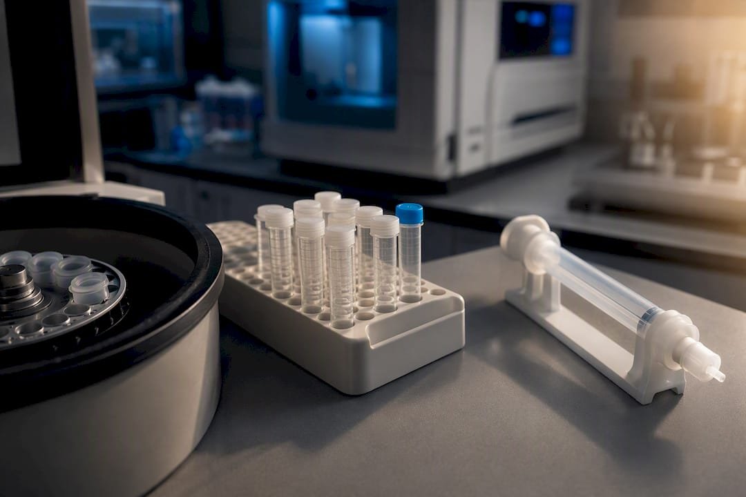
Ultracentrifugation remains the reference method most labs learned first. Differential ultracentrifugation pellets exosomes through sequential spins that clear cells, debris, and larger vesicles before a final high-speed spin (typically 100,000×g or more) isolates the vesicle-sized fraction. Density-gradient ultracentrifugation adds a separation medium such as iodixanol, sold commercially as OptiPrep, layering samples so exosomes settle at their buoyant density rather than just their sedimentation rate. This dramatically cuts protein and lipoprotein contamination but adds hours to an already long protocol and requires a fixed-angle or swinging-bucket ultracentrifuge most core facilities gatekeep by scheduling.
Size-exclusion chromatography separates by size as sample passes through a porous resin column. Commercial columns like Izon’s qEV series let smaller proteins diffuse into the resin’s pores while larger vesicles elute first, in fractions you collect and pool. SEC preserves vesicle membrane integrity better than mechanical pelleting because it avoids the shear forces of high-speed spinning.
Ultrafiltration and TFF concentrate samples across a membrane with a defined molecular weight cutoff, usually 10 to 100 DA. TFF scales this into a continuous-flow format that handles larger volumes without the membrane fouling that plagues simple centrifugal filters.
PEG precipitation works by reducing vesicle solubility so they aggregate out of solution after an incubation period, then pellet at low speed in a standard benchtop centrifuge. Kits like ExoQuick popularized this approach because it needs no specialized equipment.
Immunoaffinity capture uses antibody-coated magnetic beads targeting surface tetraspanins, most often CD9, CD63, or CD81, to pull specific vesicle populations directly from fluid. QIAGEN’s exoEasy kit applies a related membrane-affinity chemistry rather than antibody capture, binding vesicles to a spin-column membrane based on charge interactions.
Microfluidic and acoustofluidic devices are the newest entrants, separating vesicles by size, affinity, or acoustic properties within microchannels. They need only microliters of input and work well for limited clinical samples, though throughput and cross-lab standardization still lag the older methods.
Comparing Purity, Yield, and Cost Across Isolation Methods
No isolation method wins on every axis, and the comparative literature confirms it: testing precipitation, membrane affinity, SEC, density gradients, and phosphatidylserine affinity capture side by side produced meaningfully different yields and protein contamination profiles depending on which method processed the sample.
- Ultracentrifugation (density gradient): high purity, moderate yield, needs moderate sample volumes, takes multiple hours, requires a dedicated ultracentrifuge and gradient media, works well for proteomics and structural studies, costs are moderate to high once instrument time is factored in.
- SEC (qEV-type columns): high purity, moderate to good yield, works from small volumes, runs in under an hour once loaded, needs only a column and collection tubes, suits proteomics, RNA work, and functional assays, and per-sample cost sits in the moderate range once you own the equipment.
- Ultrafiltration/TFF: moderate purity, high yield, scales from milliliters to liters, processes in 30 to 90 minutes, needs a filtration membrane system, fits large-cohort screening better than single-sample precision work, and is cost-efficient at volume.
- PEG precipitation: low to moderate purity, high yield, works with small volumes (0.1 to 1 mL), finishes in 1 to 2 hours with only a benchtop centrifuge, is the cheapest option per sample, but co-isolates lipoproteins and free proteins that can skew proteomic and functional readouts.
- Immunoaffinity capture (magnetic beads): very high specificity for the targeted subpopulation, lower total yield since it selects rather than bulk-recovers, works with small volumes, takes 1 to 3 hours, needs magnetic separation racks and antibody-conjugated beads, and costs more per sample due to reagent price.
- Microfluidic devices: purity and yield vary by platform design, sample needs can be as low as 100 microliters, processing is relatively fast, but equipment access and validated protocols remain limited outside specialized labs.
The trade-off that trips up the most labs: plasma contains far more free-floating lipoproteins than cell culture medium does, and several of them overlap in size and density with exosomes. Precipitation methods concentrate this contamination right alongside your vesicles. If your downstream assay measures a protein that lipoproteins also carry, that contamination reads as a false positive. A second common error is spinning too hard for too long. Excessive g-forces during pelleting steps can rupture vesicle membranes, and once that happens, no amount of downstream cleanup recovers what you lost.
For proteomics or nanoparticle tracking analysis, prioritize SEC or a density-gradient polish step. For RNA-seq, SEC and gentle gradients again lead, since precipitation reagents can interfere with extraction chemistry. For functional assays measuring intact vesicle uptake or signaling, purity matters almost as much as viability, so avoid harsh ultracentrifugation cycles. For large-cohort biomarker screening where sample count outweighs per-sample precision, TFF or automated SEC handles volume better than any manual method.
How to Choose Your Exosome Isolation Strategy
Before picking a protocol, answer four questions honestly: What does your downstream assay actually require? How much sample do you have per subject? What equipment can you access this quarter, not eventually? And how many samples need processing per week?
- Define the downstream assay first. Proteomics and structural characterization need purity. RNA panels need RNA-compatible chemistry. Functional assays need intact, viable vesicles.
- Match sample volume to method minimums. SEC and immunoaffinity work with under a milliliter; ultracentrifugation typically wants more.
- Check equipment access realistically. A shared core ultracentrifuge booked three weeks out may force you toward SEC or precipitation regardless of theoretical preference.
- Plan your QC panel before you isolate, not after. You need a way to confirm the method worked.
- Run a small pilot batch and characterize it fully before committing a full cohort to one protocol.
The minimum QC panel worth running on any isolated exosome fraction includes particle count and size distribution by nanoparticle tracking analysis or tunable resistive pulse sensing, morphology confirmation by transmission electron microscopy, tetraspanin marker detection by western blot for CD9, CD63, and CD81, and a contamination check, typically an albumin or total protein assay, to flag lipoprotein or serum protein carryover.
Pro Tip: Run your QC panel on the first pilot batch before scaling to your full sample set. A method that looks fine on paper can produce fractions dominated by co-precipitated albumin, and you want to catch that on five samples, not fifty.
Sample Type Changes the Isolation Calculus
Plasma, cell culture medium, and urine each carry their own contamination profile, and the method that performs best in one often underperforms in another.
For blood-derived samples, use plasma rather than serum, since the clotting process in serum collection releases platelet-derived vesicles that inflate your count with material you didn’t intend to study. EDTA is the preferred anticoagulant; heparin inhibits downstream PCR reactions and should be avoided if RNA or DNA analysis follows. Process plasma promptly after draw and minimize freeze-thaw cycles, since repeated thawing degrades vesicle integrity and RNA cargo. SEC or a density-gradient step improves purity given plasma’s high lipoprotein burden.
For cell culture conditioned medium, the main contaminant is bovine-derived vesicles from fetal bovine serum. Either switch to exosome-depleted FBS well before harvest or run an extra ultracentrifugation pre-clearing step to strip serum vesicles from your base medium. A low-speed pre-clearing spin to remove cells and debris before the main isolation step is standard practice regardless of which final method you choose.
For urine and other low-protein fluids, concentration is the bigger challenge than contamination. UF/TFF or SEC handle the volume and viscosity issues reasonably well, and urine’s naturally lower protein background means precipitation methods perform better here than they do in plasma.
Getting Usable RNA Out of Isolated Exosomes
RNA integrity depends heavily on which isolation method touched your sample first. SEC and carefully run density gradients tend to preserve RNA better than PEG-based precipitation, because residual polymer from precipitation reagents can interfere with phenol-chloroform extraction and RT-qPCR chemistry downstream. If your workflow depends on precipitation for yield, validate that your specific extraction kit tolerates the carryover before committing a full study to it.
Membrane affinity spin columns designed for exosomal RNA, such as exoRNeasy, and other specialized exosomal RNA kits generally give cleaner results than adapting a generic RNA extraction protocol. Run a Bioanalyzer or equivalent trace on your first extractions, and include spike-in controls to catch inhibition before you’ve burned through your whole sample set. On the collection side, stick with EDTA plasma. Heparin’s PCR-inhibiting effect applies just as much to RNA-based assays as it does to DNA work, so the same caution belongs here.
Combining Methods When Purity Alone Isn’t Enough
Single-step isolation rarely hits both high purity and high yield, so many labs run two-step workflows instead. A fast, high-recovery first step followed by a high-resolution polish is a pragmatic compromise that balances the two when sample volume allows for it.
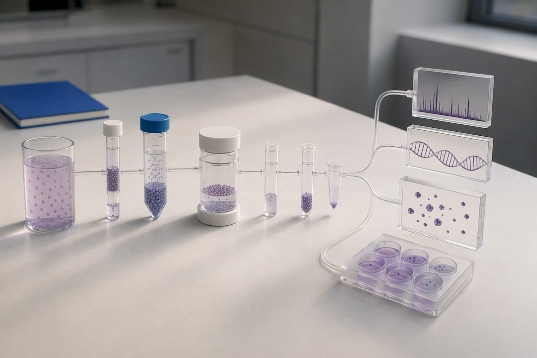
Three combinations show up most often. TFF or PEG precipitation followed by SEC gives medium-to-high purity with better throughput than SEC alone, since the first step concentrates dilute samples before the column does the fine cleanup. SEC followed by a density gradient reaches proteomics-grade purity for studies where contamination would compromise mass spectrometry results. TFF followed by automated SEC scales for biobanking and large cohorts, and automated SEC paired with tunable resistive pulse sensing adds reproducible absolute quantification without a technician manually collecting fractions for every sample.
Combinations make less sense below about half a milliliter of input, since each added step loses material and dilute samples won’t survive multiple transfers. Below that threshold, a single well-controlled step, usually SEC, is the safer default.
Where Mayflower Bioscience Fits Into Your Isolation Workflow
Mayflowerbio supports exosome research end to end, from isolation kits and reagents through detection tools for post-isolation validation. Purified exosomes are available as reference controls for method comparison studies. For labs lacking in-house nanoparticle tracking capacity, an NTA sizing service can validate particle counts and size distributions as part of routine QC without requiring instrument purchase.
A Practical Decision Framework for Exosome Isolation Methods
The conventional advice on exosome isolation treats it like a single decision: pick the “best” method and standardize on it. That framing has done real damage to reproducibility across the field. There is no best method, only the method that matches your downstream assay, and the labs that struggle most with irreproducible results are usually the ones that locked into one protocol early and never revisited it as their questions changed.
What gets underweighted is pre-analytical control. Researchers agonize over choosing between SEC and ultracentrifugation while running inconsistent blood draw timing or switching anticoagulants between cohorts. Fix your collection and storage protocol before you fix your isolation method. That single change often narrows variability more than any isolation upgrade will.
If you take one thing from this: build your QC panel before your isolation protocol, not after. Decide what “success” looks like on a particle tracker and a western blot before you run a single sample, then let that standard drive your method choice.
— Alina
Skip the Equipment Queue: Mayflower Bioscience’s Exosome Tools and Services
Waiting weeks for shared ultracentrifuge time or troubleshooting a precipitation kit that keeps co-isolating lipoproteins costs labs real momentum. Mayflowerbio carries isolation kits, purified exosome controls, and detection reagents built for the workflows described above, plus an NTA sizing service for labs that need particle validation without buying an instrument.

If you’re planning your next exosome study, here’s where to start:
- Browse exosome-related product lines for kits, controls, and detection reagents.
- Check the exosome isolation catalog for kits matched to your sample type.
- Contact technical support to discuss NTA size profiling for your current isolation protocol.
FAQ
How Do You Isolate Exosomes From Blood?
Start with plasma rather than serum, collected in EDTA tubes to avoid the platelet-derived vesicles that serum clotting releases. Process the sample quickly and choose SEC or a density-gradient step to strip out the lipoproteins plasma carries in abundance, since pre-analytical handling affects results more than most researchers expect.
How Can I Isolate RNA From Exosomes?
Use an isolation method that preserves RNA integrity, such as SEC or a careful density gradient, since precipitation reagents can leave residues that interfere with extraction chemistry. Follow with a membrane affinity RNA kit designed for exosomal RNA, such as exoRNeasy, and confirm quality with a Bioanalyzer trace before running your full sample set.
What Is the Dark Side of Exosomes?
Isolation is genuinely difficult to standardize: different methods yield different particle counts, protein profiles, and contamination levels from the same starting sample, which makes cross-study comparison unreliable without matched protocols. Lipoprotein co-isolation in plasma samples and inconsistent pre-analytical handling are the two biggest sources of misleading results in the field.
How Long Can Exosomes Be Stored?
Storage stability depends on temperature and freeze-thaw cycles rather than a fixed universal timeline, and repeated freeze-thaw degrades both vesicle structure and RNA cargo. Aliquot samples immediately after isolation and store at minus 80 degrees Celsius, minimizing the number of times any single aliquot gets thawed.
Does Mayflower Bioscience Offer Purified Exosome Controls?
Yes, Mayflowerbio supplies purified exosomes for use as reference material in method validation and comparison studies. Current pricing and specifications are listed on the product page rather than quoted here.




