

Prevent Cell Clumping in Single Cell Prep: DNase Rescue in 15–30 Min
The fastest way to prevent cell clumping is to minimize dead cells during harvest and keep your buffer cation-compatible so DNase I rescue works if you need it later. If clumps already formed, treat with 100 µg/mL DNase I plus 5 mM MgCl2 for 15 to 30 minutes at room temperature. Prevention beats rescue almost every time, but when DNA-mediated aggregation hits, DNase I is your most reliable fix. Additives like Pluronic F-68 help with mechanical protection, but validate them against your specific assay before you commit a full run.
TL;DR:
- Removing dead cells during harvest prevents most DNA-driven clumping and reduces reliance on DNase I rescue.
- Using buffers with magnesium and avoiding EDTA at the cleaning stage ensures DNase activity remains effective if needed.
- DNase I treatment at 100 µg/mL with MgCl2 for 15 to 30 minutes can break up existing aggregates, but actin from dead cells can inhibit its effectiveness.
- Additives like Pluronic F-68 protect cells from shear but may alter mechanical properties, so validate in your specific assay first.
- Mechanical steps like gentle pipetting and filtering only at the end help prevent cell lysis and subsequent DNA release.
Table of Contents
- What Causes Cell Clumping?
- How Do You Prevent Cell Clumping Before It Starts?
- What Do You Do When Cells Are Already Clumped?
- Anti-Aggregation Additives: What Helps and What They Cost You
- Quick Checklist Before Counting, Sorting, or Loading a Microfluidic Chip
- What Researchers Get Wrong About Clump Rescue
- Mayflower Bioscience Resources for Cell Separation and Sample Prep
- Get the Right Tools for Clean Single-Cell Prep
- Authoritative Protocols and Studies
- Sources
- FAQ
What Causes Cell Clumping?
Free DNA from lysed or dying cells is the single most common driver of aggregation. Dead cells rupture and release DNA that is genuinely sticky and viscous, and that viscosity binds otherwise healthy cells together into clumps that clog cytometers and choke microfluidic channels, according to flow cytometry guidance from the University of Florida.
Several upstream mistakes feed this problem before you ever reach the bench:
- Overdigestion with proteases during tissue dissociation, which damages membranes and triggers cell death
- Freeze/thaw cycles that rupture a fraction of the population every time
- Mechanical stress from vigorous vortexing or narrow-bore pipetting
- Overconfluent cultures harvested too late, when density itself stresses cells
- Contamination or residual debris carried over from prior wash steps
Once DNA is in solution, it does not just make cells stick to each other. It fouls microfluidic surfaces and drags dead-cell fragments into the same aggregate as viable cells, which throws off both counting accuracy and sort purity.
How Do You Prevent Cell Clumping Before It Starts?
Most clumping problems trace back to decisions made during harvest and wash, not to some mysterious property of the cells themselves. Get the basics right at that stage and you avoid most rescue work entirely.
- Choose the right buffer. Use PBS (1X) with 0.04% to 1% BSA for most single-cell preps, or up to 10% FBS when the cell type needs extra viability support. Switch to full culture media instead of PBS when cells are fragile or long processing times are unavoidable.
- Watch your cations. Avoid EDTA in any buffer you might later need for DNase rescue, since EDTA chelates the magnesium DNase requires functioning. If you anticipate needing DNase, keep Mg2+ available from the start.
- Handle gently. Use wide-bore pipette tips, mix with 10 to 15 gentle strokes, and skip vigorous vortexing entirely. Every extra minute between prep and assay is another minute for cells to die and release DNA.
- Control concentration and spin speed. Keep cells at the concentration your protocol specifies, and centrifuge at the gentlest speed that still pellets the sample. Excess shear during centrifugation is a quiet, frequently overlooked source of lysis.
- Inspect before you proceed. Reserve your supernatant until you have confirmed the pellet looks clean, then do a quick visual check under the microscope for stringy or viscous material before moving to counting.
Institutional flow cytometry guidance consistently frames prevention as faster and less destructive than rescue. Repeated harsh filtration to fix a clumpy sample after the fact costs you cells you can never get back.
Pro Tip: Keep a small aliquot of your wash buffer with Mg2+ on hand even if your main protocol uses EDTA elsewhere. If clumping shows up mid-run, you will not have time to reformulate a buffer from scratch.
For a broader look at sample handling across cell biology workflows, Mayflowerbio’s cell biology research tips cover dead-cell reduction and viability checks in more depth.
What Do You Do When Cells Are Already Clumped?
DNase I is the standard rescue for DNA-mediated clumps, and it works because it degrades the sticky extracellular DNA holding the aggregate together rather than trying to force cells apart mechanically.
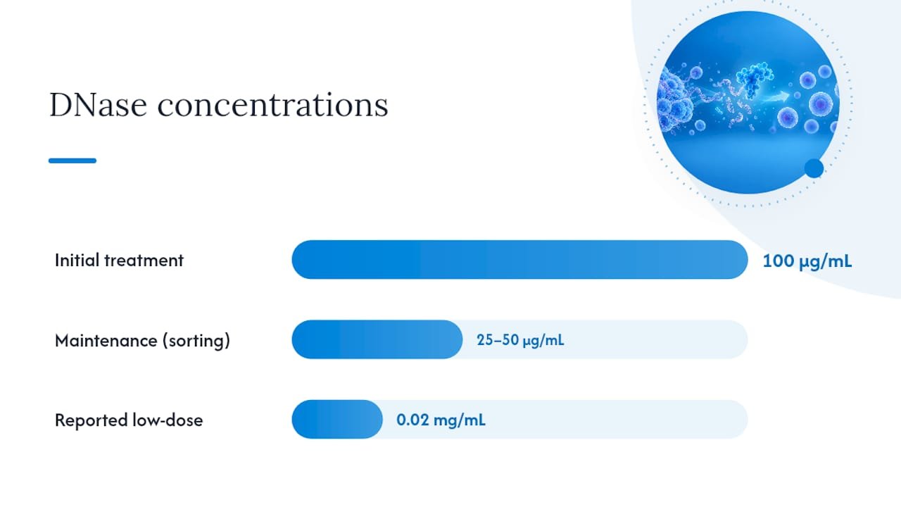
- Treat with 100 µg/mL DNase I plus 5 mM MgCl2 in HBSS for 15 to 30 minutes at room temperature, then wash in a buffer that still contains Mg2+, following the DNase and clump rescue protocol from UPF.
- During active sorting, a maintenance concentration of 25 to 50 µg/mL DNase is commonly used to keep clumps from re-forming as the run continues.
- Some labs get equivalent results with as little as 0.02 mg/mL for specific cell types, so treat the standard concentration as a starting point rather than a fixed rule.
- Never add EDTA to a buffer you plan to treat with DNase. It will chelate the magnesium the enzyme needs and the treatment will simply fail.
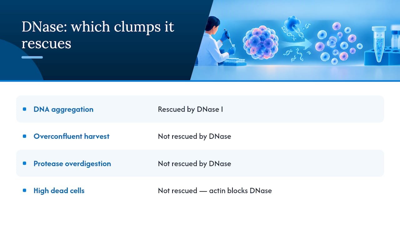
There is a catch worth knowing before you rely on DNase as your fallback: actin released from dying cells can irreversibly inhibit DNase I activity. Samples with a very high dead-cell burden may not respond to treatment at all, which is exactly why prevention has to come first, according to University of Florida flow cytometry guidance.
If DNase alone does not resolve the aggregate, wide-bore pipette trituration is the next safest mechanical step. Filtering through a 30 to 70 µm strainer helps, but Illumina’s sample prep guidance notes that forcing large aggregates through a filter can cost up to 30% cell loss, so filter gently and only at the last wash. Before committing your whole sample to an assay, re-count, check viability against a 70% or higher target for most single-cell protocols, and test a small aliquot first.
Anti-Aggregation Additives: What Helps and What They Cost You
Pluronic F-68 is the most widely used additive for long sorts and bioreactor work, and it earns that reputation by reducing cell attachment to surfaces and protecting against shear and bubble damage during extended processing.
That protection comes with a caveat. Multiple studies document that Pluronic can alter cell mechanical properties and may stiffen certain cell types even at low concentrations, according to research on Pluronic F-68 effects on mammalian cells. If your downstream assay depends on label-free or mechanical readouts, that stiffening can quietly skew your results.
- Pluronic F-68 / poloxamers: Strong protection for long sorts; test carefully before use in mechanotype-sensitive assays.
- PVA plus dextran sulfate: Effective for controlling aggregate size in suspension culture, including hPSC expansion work where it preserves pluripotency while limiting excess clustering.
- Substrate interactions: Poloxamer additives can reduce protein fouling on PDMS and glass microfluidic surfaces, but they may need extra wash steps to avoid interfering with enzymatic chemistries downstream.
Pro Tip: If you run label-free or mechanical assays, run a side-by-side comparison with and without your chosen additive before trusting the data. A small stiffening effect can look like a biological result if you are not watching for it.
Quick Checklist Before Counting, Sorting, or Loading a Microfluidic Chip
Run this sequence right before you commit a sample to any downstream instrument:
- Visual check for stringy or viscous material in the tube.
- Gentle pipette mixing, 10 to 15 strokes with a wide-bore tip.
- DNase rescue if viscosity is present, 15 to 30 minutes at room temperature.
- Filter at the last wash step only, using a 30 to 70 µm strainer, if aggregates persist.
- Re-count and check viability before proceeding.
| Decision | Criteria |
|---|---|
| Proceed | Viability ≥70%, minimal debris, no visible stringiness |
| Reprocess | Viability borderline, some debris, clumps resolve after DNase |
| Discard | High dead-cell burden, DNase fails to resolve clumps, contamination suspected |
Budget 15 to 30 minutes for DNase incubation, a few minutes per wash cycle, and roughly 10 minutes for a proper re-count and viability check before you load your instrument.
What Researchers Get Wrong About Clump Rescue
Most troubleshooting guides treat DNase I like a universal fix, and that framing sets researchers up to waste time. It is a strong rescue for DNA-mediated aggregation specifically. It does nothing for clumps caused by overconfluent harvests, protease overdigestion, or genuinely high dead-cell fractions where actin inhibition blocks the enzyme outright.
The bigger blind spot is additive selection. Labs reach for Pluronic F-68 because it is the default answer for shear protection, then run label-free or mechanical assays on the same sample without checking whether the additive shifted cell stiffness. That is not a hypothetical risk. It is a documented effect in the literature, and it means the additive that saved your cells during a long sort might be quietly corrupting your downstream readout. Validate per assay, every time, rather than assuming a protective additive is a neutral one.
If there is one habit worth building, it is treating your dead-cell percentage as the leading indicator, not an afterthought you check after something goes wrong. A sample that starts clean rarely needs rescue at all.
— Alina
Mayflower Bioscience Resources for Cell Separation and Sample Prep
Mayflowerbio is dedicated to providing innovative products, tools, and solutions for scientific research in drug discovery, cell biology, and molecular biology, backed by US-based customer support. Our specialized lines include multiplex assays, antibodies, cell counting slides, infectious disease reagents, and cell separation solutions built for exactly the kind of aggregation problems covered here.
[Alina’s scientific background and credentials to be inserted here.] If you are validating a DNase protocol for a nonstandard cell type or need bulk reagent sourcing, contact Mayflowerbio’s technical team directly for protocol guidance and supply support.
Get the Right Tools for Clean Single-Cell Prep
Prevention and rescue both depend on having the right reagents and counting tools on hand, not just the right protocol.

A generic reagent catalog does not tell you which buffer additive is compatible with your microfluidic chip or your assay’s mechanotype sensitivity. Mayflowerbio’s cell separation category is built specifically for researchers troubleshooting aggregation in single-cell workflows, and pairing it with cell counting tools lets you verify viability and concentration before you commit a full sample to a sort or assay. Our US-based technical support team can help you validate DNase concentrations and buffer formulations for your specific cell type before you burn a sample on a bad run. Browse the full cell biology product line to find the reagents and tools that fit your prep, or reach out directly for bulk procurement pricing and protocol support.
Authoritative Protocols and Studies
- Protocol digest on DNase and clump rescue (UPF)
- 10x Genomics cell preparation guidance for single-cell protocols
- University of Iowa: disaggregation of clumped cells
- Pluronic F-68 effects on mammalian cells
This article is general information, not a substitute for advice from a qualified doctor. Consult a qualified healthcare professional about your own circumstances before acting on anything here.
Sources
- Protocol digest (DNase and clump rescue) — UPF
- Review / study on effects of Pluronic F‑68 on mammalian cells (PMC6404947)
FAQ
What Causes Cells to Clump?
Cell clumping is usually caused by free DNA released from dead or dying cells, which is sticky enough to bind otherwise healthy cells together. Overdigestion, freeze/thaw cycles, mechanical stress, overconfluent harvests, and residual debris all increase the dead-cell fraction that drives this, according to UF flow cytometry guidance.
What Term Describes Cell Clumping?
Researchers typically call this “cell aggregation” or “cell clumping” in flow cytometry and single-cell contexts, and the underlying phenomenon is often described as DNA-mediated aggregation when free extracellular DNA is the cause. The term applies broadly across sorting, counting, and microfluidic sample prep.
What Is Blood Cell Clumping?
In hematology, blood cell clumping usually refers to red or white blood cells sticking together in a sample, often from anticoagulant issues, cold agglutinins, or cell damage during collection. The mechanism overlaps with general cell aggregation. Free DNA and debris from lysed cells still play a major role.
What Does Clumping Mean in Biology?
Clumping in biology describes cells adhering to each other in suspension rather than remaining as discrete single cells, which interferes with accurate counting, sorting, and microfluidic processing. It results from sticky extracellular material, most commonly DNA from dead cells, binding viable cells together into aggregates.
Does DNase I Always Fix Clumped Samples?
No. DNase I resolves DNA-mediated clumping effectively when given adequate Mg2+ and correct incubation time, per the UPF rescue protocol. Samples with a very high dead-cell burden can resist treatment because actin released from dying cells inhibits the enzyme, which is why minimizing dead cells during harvest remains the more reliable long-term fix.




