

Growth Factor Selection for Labs: Match Receptors, Read CoAs, Cut Cost
Choose the growth factor that matches your target cell’s receptor-signaling profile, then verify the supplier’s bioactivity data and lot traceability before you scale. Shortlist two or three candidate families, request a certificate of analysis with bioactivity assay details, and run a small titration before committing to a protocol. The trust anchors worth checking every time are bioactivity potency, endotoxin level, host-cell origin, and lot number.
TL;DR:
- Ensure growth factors are sourced from suppliers providing detailed bioactivity units, lot traceability, and animal-free or carrier-free formulations if required by your protocol or compliance needs.
- Confirm your target cells express the specific receptors for the chosen growth factor, as receptor presence determines responsiveness regardless of growth factor reputation.
- Run a small titration and check a new lot’s bioactivity with a defined assay before scaling up, especially if the lot lacks validated potency data.
- For regulated or clinical applications, qualify growth factors with documented origin, endotoxin levels, residual DNA, and sterility, and perform comparability studies when switching lots or suppliers.
- Prioritize high-purity, validated growth factors for sensitive work, but consider testing for cost-effectiveness with in-house titrations during early discovery phases.
Table of Contents
- Common growth factor families and cell-type matchups
- Choosing source and formulation: recombinant versus animal-derived, carrier-free versus carrier-protein
- What to read on the datasheet and CoA: an itemized checklist
- Handling, reconstitution, storage, and dosing best practices
- Optimizing concentrations and combinations: DoE, Bayesian optimization, and RSM workflows
- Regulatory and qualification considerations for clinical or GMP-adjacent work
- Procurement checklist and supplier workflow
- Balancing performance and cost in growth-factor selection
- How Mayflower Bioscience can help
- Sources
- FAQ
Common growth factor families and cell-type matchups
Selecting the right family starts with the biological question you’re asking. Epidermal growth factor (EGF) drives epithelial proliferation and is a mainstay in keratinocyte and organoid culture. Fibroblast growth factor 2 (FGF2, also called bFGF) supports self-renewal in stem cell culture and proliferation in fibroblasts and mesenchymal lines. Platelet-derived growth factor (PDGF) promotes proliferation in fibroblasts, smooth muscle, and glial cells, while insulin-like growth factor (IGF) supports survival and metabolic signaling across many cell types. Transforming growth factor beta (TGF-β) isoforms regulate differentiation and can either promote or suppress proliferation depending on context, and vascular endothelial growth factor (VEGF) is the standard choice for angiogenesis assays and endothelial cell maintenance. Colony-stimulating factors and select interleukins round out the panel for hematopoietic and immune cell work.
Receptor expression on your target cells, not the growth factor’s general reputation, is what determines whether it will do anything useful. A cell line lacking the cognate receptor will not respond regardless of concentration, since receptor-signaling context determines specificity through pathways like MAPK/ERK and PI3K/AKT. Before ordering, check the literature or expression datasets for your cell type to confirm the receptor is actually present.
- EGF suits epithelial and keratinocyte proliferation assays.
- FGF2/bFGF suits stem cell self-renewal and fibroblast proliferation.
- PDGF suits fibroblast, smooth muscle, and glial proliferation.
- VEGF suits angiogenesis and endothelial maintenance assays.
- TGF-β isoforms suit differentiation studies where proliferation control matters.
Starting concentrations vary widely by application, and product-specific datasheets (often echoing ATCC or kit protocols) remain the most reliable starting point rather than a generic industry figure.
Choosing source and formulation: recombinant versus animal-derived, carrier-free versus carrier-protein
Expression host shapes more than yield. Mammalian systems produce post-translational modifications closest to native human protein, bacterial expression is cheaper but can miss glycosylation that some factors need for full activity, and yeast or insect systems sit somewhere in between. A generalized recombinant expression workflow shows that FGF, IGF, PDGF, and TGF-β family members can each be produced with workable bacterial protocols, but host choice and post-translational needs differ by family, so the “best” host is protein-specific.
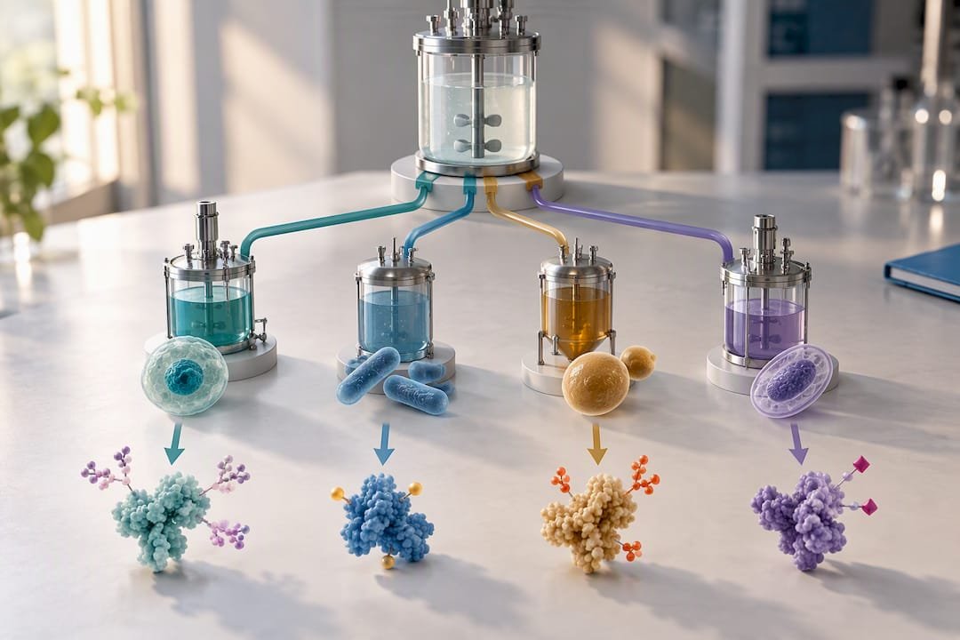
Animal-derived growth factors carry batch-to-batch variability and adventitious-agent risk that most regulated or reproducibility-sensitive workflows try to avoid, which is why non-animal, recombinant sources are the default recommendation for anything moving toward scale or compliance. Carrier proteins like bovine serum albumin stabilize dilute stock solutions but can interfere with assays sensitive to exogenous protein, so carrier-free formulations are preferable when your downstream assay measures total protein or uses mass spectrometry.
Ask suppliers for these details before you buy:
- A clear origin statement (species, expression host, recombinant versus native).
- Whether the formulation includes a carrier protein and which one.
- The exact formulation buffer and any excipients.
Pro Tip: Request the animal-free version of a growth factor when your protocol or funder requires xeno-free documentation, even if the standard version is cheaper. Suppliers such as Mayflowerbio list animal-free recombinant FGF-21 and animal-free TGF-α with origin statements included on the product page.
What to read on the datasheet and CoA: an itemized checklist
A certificate of analysis is only useful if you know which fields actually matter. Run through this list on every new lot:
- Bioactivity assay and units. Confirm the assay type (proliferation, receptor binding) and the ED50 or unit definition, since mass alone does not tell you potency.
- Purity. Look for SDS-PAGE or HPLC purity above the threshold your application requires.
- Endotoxin level. Expressed in EU per microgram, this matters most for cell-based assays sensitive to inflammatory activation.
- Host-cell protein and residual DNA. Relevant for any downstream regulated or GMP-adjacent use.
- Sterility and formulation. Confirms the buffer, carrier presence, and fill format match what you expect.
- Lot number and expiry. Essential for traceability and for correlating any experimental drift back to a specific batch.
Mass-based dosing (micrograms per milliliter) and activity-based dosing (units per milliliter) are not interchangeable, since two lots with identical mass concentration can differ in potency. If a CoA lacks bioactivity data entirely, treat that as a red flag and ask the supplier for reference-lot potency data or run a small-scale potency check yourself before committing to a full experiment.
Handling, reconstitution, storage, and dosing best practices
Reconstitution technique affects activity as much as the protein itself. Most lyophilized growth factors reconstitute in sterile water or a dilute acid or base buffer specified on the datasheet, and many benefit from a carrier protein added to the working stock (not necessarily the lyophilized powder) to prevent surface adsorption losses at low concentration.
- Aliquot immediately after reconstitution to avoid repeated freeze-thaw cycles, which degrade activity.
- Store lyophilized powder at negative 20 degrees Celsius or lower and reconstituted stock per the datasheet, typically negative 20 to negative 80 degrees Celsius for long-term storage.
- Titrate a new lot across a concentration range around the supplier’s suggested starting point rather than assuming the listed value transfers directly to your system.
- Add heparin or heparan sulfate for FGF family members, since these factors depend on heparan sulfate proteoglycans for stable receptor engagement.
Pro Tip: Keep a simple lot logbook that records reconstitution date, buffer, storage location, and freeze-thaw count for every aliquot, and retain a small frozen sample from each lot for later comparison.
Optimizing concentrations and combinations: DoE, Bayesian optimization, and RSM workflows
One-factor-at-a-time (OFAT) testing wastes reagent and misses interaction effects between growth factors that act on overlapping pathways. Design of experiments (DoE), Bayesian optimization, and response surface methodology (RSM) address this by modeling multiple factors simultaneously.
- Screen broadly first. Use a fractional factorial or Latin hypercube design to test several growth factors and concentrations at once rather than one at a time.
- Build a model. Fit a response surface or Gaussian process model to the screening data to identify which factors and interactions matter most.
- Optimize under constraints. Use the model to propose the next experiments, weighting toward combinations likely to improve your readout while respecting cost limits on expensive factors.
- Validate. Confirm the model’s top candidate combination with independent replicates before adopting it as standard protocol.
Active learning and Bayesian optimization workflows have been shown to identify optimized media and cytokine combinations with far fewer experiments than traditional one-factor-at-a-time approaches, which matters directly for growth factor budgets since these reagents often dominate serum-free media cost.
Design choices that determine whether your optimization is trustworthy include the readout you’re modeling (viability, marker expression, yield), replicate count per condition, and awareness of batch effects between reagent lots run on different days. To control cost, minimize use of the most expensive factors in early screening rounds and consider immobilization or tethering approaches, which can reduce soluble consumption while preserving local bioactivity in adherent or scaffold-based systems, as described in work on reducing growth-factor consumption.
Regulatory and qualification considerations for clinical or GMP-adjacent work
Growth factors used in manufacturing cell or gene therapies are classified as ancillary materials, and qualifying them properly is not optional once you move past basic research. ISO 20399 sets out ancillary-material guidance recognized by regulatory programs, covering reagents and growth factors used in cell processing specifically.
FDA CMC guidance for gene and cell therapy recommends qualifying reagents used during manufacture, including growth factors, with documented testing to reduce adventitious-agent risk and process variability, generally favoring clinical-grade or well-characterized materials over standard research-grade stock.
Supplier qualification for this tier of work should include:
- Documented origin and manufacturing history for each lot.
- Endotoxin and sterility testing results.
- Residual host-cell DNA and protein data.
- Retained samples for future comparability testing.
When you switch lots or suppliers, plan a comparability study rather than assuming equivalence. Small formulation or potency differences between lots can shift downstream cell behavior in ways that are easy to miss until they show up in a failed batch.
Procurement checklist and supplier workflow
Before placing an order, confirm the supplier can provide a full CoA with bioactivity data, an origin statement, and lot-specific documentation on request. A short technical exchange upfront saves far more time than troubleshooting an underperforming lot later.
- Request the bioactivity assay type and units used to establish potency.
- Ask whether animal-free or carrier-free versions exist for your application.
- Confirm expected turnaround for CoA and technical documentation requests.
- Ask about lot-to-lot consistency data if you plan to scale.
Some suppliers provide recombinant growth factors including animal-free options across several families, with technical support and CoAs available on request, alongside broader catalogs of cell biology reagents and bioassay services for labs that need testing support beyond the reagent itself.
Balancing performance and cost in growth-factor selection
Pay for validated, high-purity bioactivity data when the work feeds a regulated pipeline or a result you plan to publish. During early discovery, it’s reasonable to optimize for cost and run your own in-house potency check before scaling, since a modest titration catches most underperforming lots before they waste a full experiment.
— Alina
How Mayflower Bioscience can help

Sourcing a growth factor is only half the procurement problem. The harder half is getting a supplier who will actually hand over bioactivity data, origin statements, and lot documentation without a week of back-and-forth email. Mayflower Bioscience carries recombinant growth factors, including animal-free formulations such as FGF-2 (animal free), rat recombinant, with technical support based in the United States and CoAs available on request for the lot you’re actually buying, not a representative sample lot.
If your protocol calls for a specific family or ortholog, browse the full growth factors catalog or send us the receptor-signaling profile and endpoint you’re targeting, and our technical team can help you narrow the shortlist before you commit reagent budget to a titration series.
Sources
- CBER Standards Recognition Program for Regenerative Medicine Therapies (ISO 20399 recognition document)
- Growth factor signaling and cellular responses: receptor expression guides functional outcomes
FAQ
What are the four main types of growth factors?
Growth factor families are typically grouped by signaling role rather than a fixed list of four, but the most commonly referenced groups are epidermal growth factor (EGF), fibroblast growth factor (FGF), platelet-derived growth factor (PDGF), and transforming growth factor beta (TGF-β). Vascular endothelial growth factor (VEGF) and insulin-like growth factor (IGF) are also core families depending on the application.
Which growth factor is best?
There is no single best growth factor since the right choice depends entirely on which receptors your target cells express and which endpoint you’re measuring. Receptor-signaling context determines whether a given factor will produce a useful response, so selection starts with confirming receptor expression, not with a general ranking.
Does growth factor serum actually work?
Serum and defined growth factor supplements both support cell proliferation, but serum introduces lot-to-lot variability that defined, recombinant growth factors are specifically meant to reduce. For reproducibility-sensitive or regulated work, well-characterized recombinant growth factors with documented bioactivity are generally preferred over undefined serum supplementation.
How to determine the growth factor?
Start by confirming which receptors your target cell type expresses for the endpoint you want, whether that’s proliferation, differentiation, or survival, then shortlist candidate families accordingly. From there, request bioactivity and CoA data from suppliers and run a small titration to confirm the working concentration performs as expected in your own system.




