

Fix Lentiviral Transduction in Primary and Suspension Cells: 0.1–10 MOI
The fastest path to better transduction is a bundle, not a single trick: confirm a high functional titer, put cells in the right activation state, add a compatible enhancer, and run a small MOI matrix before committing to a large experiment. No single lever fixes a low-efficiency system on its own. Treat the first optimization round as a diagnostic exercise, and confirm your institutional biosafety approvals are in place before handling concentrated virus.
TL;DR:
- Achieving high transduction efficiency requires optimizing multiple factors such as virus titer, cell activation state, and enhancer use, with no single trick providing a complete fix.
- Using gentler concentration methods like lower-speed ultracentrifugation or ultrafiltration improves virus recovery and transduction, especially in primary T cells, by reducing virion damage.
- Spinoculation combined with retronectin coating significantly enhances transduction rates but may increase cytotoxicity, so viability monitoring and protocol adjustments are necessary.
- Soluble enhancers like poloxamer-based formulations or Vectofusin-1 offer better viability and transduction results in hard-to-transduce cells, with trade-offs in cost and cell type compatibility.
- Proper cell activation, medium composition, and timing greatly influence efficiency, making it critical to record conditions precisely and run dose-response and time-course experiments for optimization.
Table of Contents
- How to improve virus stock quality and measure functional titer
- Using spinoculation and retronectin to increase contact and capture
- Soluble enhancers: polybrene, LentiBOOST, and Vectofusin-1 dosing and trade-offs
- Optimize target cell state: activation, medium, and timing
- Designing an MOI and optimization matrix: controls and time points
- Common troubleshooting steps and quick checks
- Selection of target cell types and culture conditions
- Post-transduction selection and expansion methods
- Assessment of transduction efficiency and gene expression stability
- Quality control of lentiviral preparations: purity and endotoxin levels
- Balancing high transduction with downstream quality: a practitioner’s perspective
- Where to find lentiviral supplies and technical support
- Authoritative protocols and biosafety guidance
- Sources
- FAQ
How to improve virus stock quality and measure functional titer
Functional titer, expressed as transducing units per milliliter (TU/ml), is the number that predicts what will happen in your experiment. p24 antigen ELISA and RNA-based quantification are useful quality checks during production, but they measure particle mass or genome copies, not infectivity, so a stock can look strong on paper and still transduce poorly.
Concentration method matters as much as the titration assay. Ultracentrifugation at very high speeds is common, but reducing centrifuge speed to roughly 20,000 g has been shown to improve viral recovery and correlate with higher transduction in primary CD45RA+ T cells compared with the more common 90,000 g protocol, likely because gentler spins cause less physical damage to virions. Ultrafiltration is a reasonable alternative when a centrifuge with the right rotor is not available.
Producer-cell health drives everything downstream. Low-passage, actively growing HEK293T cells and well-optimized plasmid ratios during transfection change functional titer substantially, independent of any downstream concentration step.
- Store concentrated virus in single-use aliquots at negative 80 degrees Celsius.
- Avoid repeated freeze-thaw cycles: titer measurably declines after the third cycle, so plan aliquot volumes around actual experiment size rather than convenience.
Using spinoculation and retronectin to increase contact and capture
Spinoculation increases the physical contact between virus and cell by centrifuging the mixture in a small volume, and it is often paired with retronectin, a fibronectin fragment that coats the culture surface and captures both virus and cells in proximity. In a CAR T-cell study, spinoculation combined with retronectin produced roughly 63% transduction compared with about 10% for polybrene alone, while polybrene and spinoculation with polybrene both increased cytotoxicity at higher concentrations.
- Coat non-tissue-culture-treated plates with retronectin solution and incubate at 4 degrees Celsius overnight or at room temperature for a few hours, then block and wash before adding virus.
- Concentrate virus and cells into the smallest practical volume to maximize particle-to-cell contact.
- Centrifuge at a moderate speed for 60 to 90 minutes rather than defaulting to a single fixed protocol; fragile primary cells often tolerate shorter spins better.
- Monitor viability at 24 and 48 hours post-spin, since increased cytotoxicity is the main trade-off with this method.
Spinoculation is worth avoiding, or using at reduced speed and time, with cells known to be shear-sensitive, such as some stem and progenitor populations.
Pro Tip: Run one well with retronectin and no spin alongside your spinoculated well: the comparison tells you how much of the benefit is coming from surface capture versus centrifugal force.
Soluble enhancers: polybrene, LentiBOOST, and Vectofusin-1 dosing and trade-offs
Polybrene is the oldest and cheapest soluble enhancer, typically used in the low micrograms-per-milliliter range, but it raises cytotoxicity risk in primary and sensitive cell types and should be kept to short exposure windows when used at all.
Poloxamer-based enhancers, sold under names like LentiBOOST and built on Synperonic F108 chemistry, offer a gentler alternative. In cord-blood CD8+ T cells, poloxamer-based enhancement increased both transduction and vector copy number, especially at low MOI, with minimal impact on viability, and GMP-grade formulations of this enhancer class are already used in clinical manufacturing.
Vectofusin-1 is a soluble peptide enhancer that works across multiple pseudotypes and fits well into closed, automated bioprocessing platforms, making it a practical choice for labs scaling toward clinical-grade or high-throughput workflows rather than small benchtop experiments.
- Polybrene: lowest cost, highest toxicity risk; best for robust cell lines, not primary lymphocytes.
- Poloxamer (LentiBOOST class): favors low-MOI experiments where virus is limiting and viability matters.
- Vectofusin-1: favors pseudotype flexibility and closed-system compatibility.
Poloxamer-based enhancers improve transduction in hard-to-transduce cells with only a modest effect on viability, which is why they show up repeatedly in translational and clinical-scale protocols.
Optimize target cell state: activation, medium, and timing
Cell state at the moment of exposure often matters more than any reagent choice. For T cells, transduction efficiency is commonly highest 24 to 48 hours after anti-CD3/CD28 activation, when the cells are cycling but not yet exhausted. Checking activation markers like CD25 and CD45RO alongside your transduction readout tells you whether a low percentage reflects poor virus entry or simply an under-activated population.
Medium composition is not a side detail. Human serum, fetal calf serum, and specific cytokine cocktails all shift how permissive cells are to lentiviral entry, so any optimization log should record exactly what medium and supplements were used, since a protocol that works with one serum source may not transfer cleanly to another lab’s conditions.
- Time transduction to the activation window, not to a fixed day after thaw.
- Record serum type, cytokines, and concentration for every condition tested.
- Measure functional outcomes, such as cytotoxic activity or persistence, not only the percentage of reporter-positive cells, since stronger activation can also push cells toward a different differentiation state.
Designing an MOI and optimization matrix: controls and time points
A single MOI test tells you almost nothing about where your bottleneck sits. A practical optimization workflow runs a small dose series with parallel viability and reporter readouts across multiple time points, rather than escalating a single condition.
- Test an MOI series starting around 0.1, 0.5, 1, 5, and 10, using your functional titer, not particle count, to calculate each dose.
- Include enhancer and no-enhancer arms at each MOI, plus a mock (no virus) and virus-only control.
- Read out reporter-positive percentage and viability at more than one time point, typically 48 and 96 hours, to separate transient toxicity from stable integration.
- Confirm integration or transgene function with an orthogonal assay, such as ddPCR for vector copy number, when the reporter signal alone is ambiguous.
This structure isolates whether a poor result comes from entry, expression, or cell health, instead of leaving you to guess.
Common troubleshooting steps and quick checks
Low transduction has two broad causes, and separating them changes what you do next. Low infectivity points to the virus stock itself: re-titer it, and check whether freeze-thaw cycling or a harsh concentration step damaged particles. Low permissiveness points to the cells: revisit activation timing, medium, and enhancer choice before touching the virus again.
A few physical details get overlooked often enough to be worth a standing checklist:
- Filter virus-containing supernatant through 0.45 micrometer, not 0.22 micrometer, filters, since the tighter pore size can strip out a meaningful fraction of particles.
- Track producer-cell passage number; late-passage HEK293T cultures produce measurably lower titers even under an unchanged protocol.
- If an enhancer appears toxic, try a shorter exposure window or a lower concentration before abandoning the enhancer class entirely.
Pro Tip: Keep a single shared log of filter type, centrifugation history, and producer passage number across your team: most “the protocol stopped working” problems trace back to one of these three variables changing quietly.
Selection of target cell types and culture conditions
Not every cell type responds to the same optimization stack, and matching the approach to the biology saves time. Adherent lines like HEK293T or HeLa are typically easy to transduce and tolerate polybrene and standard spinoculation well, making them useful for initial virus QC before moving to a harder target.
Primary T cells sit in the middle: they respond well to retronectin, spinoculation, and poloxamer-based enhancers, but their susceptibility window is tied tightly to activation state and culture medium, as covered above.
Suspension and stem or progenitor cells are the hardest group. Hematopoietic stem cells (HSCs) are largely quiescent, and quiescence itself limits transduction independent of virus quality or enhancer choice. Entry restriction and metabolic constraints, including limited pyrimidine pools, can be the actual bottleneck in these cells, and experimental combinations such as cyclosporin H with deoxynucleosides have been reported to overcome some of these restrictions, though such approaches require institutional review before use.
Culture conditions should be chosen with the downstream assay in mind rather than defaulting to whatever medium a protocol paper used. Cytokine support that keeps HSCs cycling enough to be transducible can also push them toward differentiation, so a culture condition that maximizes transduction is not automatically the one that preserves the cell population you actually need.
Post-transduction selection and expansion methods
Once transduction is complete, the goal shifts from maximizing entry to isolating and expanding a clean, representative population of transduced cells without distorting the underlying biology.
Antibiotic selection, using resistance genes like puromycin or blasticidin built into the vector, works well for cell lines but is rarely appropriate for primary cells, where the added stress can compound transduction-related toxicity. Fluorescence-based sorting, using a reporter gene or a surface marker tag, is gentler and lets you confirm the sorted population’s purity directly rather than inferring it from drug survival.
Expansion protocols should track growth kinetics against an untransduced control from the same donor or passage. A transduced population that grows noticeably slower than its control is telling you something about vector burden or off-target toxicity, not just about transduction efficiency.
- Sort or select as early as practical after transduction to avoid outgrowth of a non-representative subpopulation.
- Expand in the same medium and cytokine conditions used during transduction unless a specific reason requires a change.
- Re-check viability and phenotype markers partway through expansion, not only at the final harvest point.
Cryopreserving an aliquot of the expanded, selected population immediately after expansion, rather than only at the end of a longer downstream process, protects the work if a later assay reveals a problem that traces back to this stage.
Assessment of transduction efficiency and gene expression stability
Percentage of reporter-positive cells by flow cytometry is the most common efficiency readout, and it is a reasonable first-pass number, but it does not tell you how many vector copies each cell carries or whether expression will hold up over time.
Vector copy number (VCN), measured by ddPCR, adds that missing dimension. ddPCR-based infectious titer and VCN assays are generally within about 30% of flow cytometry-based functional titers and can reliably determine copy number for cell products where regulatory or translational thresholds apply. A high reporter percentage paired with an unexpectedly high VCN can indicate multiple integration events per cell, which matters for both safety and manufacturing consistency.
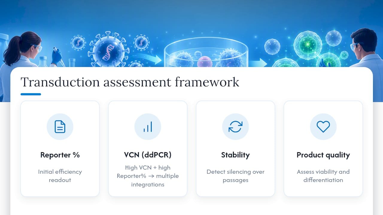
Stability is a separate question from initial efficiency. Silencing of transgene expression over serial passages is a known issue with some promoter and vector combinations, so a single efficiency measurement taken shortly after transduction says little about expression a few weeks later. Reassessing reporter-positive percentage and, where relevant, functional protein output at a later passage catches silencing before it affects a larger experiment or a banked cell product.
Different enhancers can produce comparable reporter percentages while differing in downstream viability, differentiation, or memory phenotype, so efficiency and product quality should be evaluated together rather than treating a single transduction percentage as the full picture.
Quality control of lentiviral preparations: purity and endotoxin levels
A titer number alone does not tell you whether a virus preparation is clean enough to trust in a sensitive assay or a translational protocol. Purity and endotoxin testing round out the QC picture that functional titration starts.
Residual host-cell protein and plasmid DNA carryover from producer cells can affect downstream cell health independent of the virus itself, which is one reason two preparations with identical TU/ml can behave differently in the same cell type. Endotoxin contamination, typically measured by a limulus amebocyte lysate (LAL) assay, is a standard release criterion for any preparation destined for primary cells or translational use, since even low-level endotoxin can activate immune cells and confound a transduction readout.
A practical QC panel for a lentiviral prep includes functional titer by TU/ml or ddPCR, endotoxin level, and a sterility check, run before the stock is used in a sensitive experiment rather than after a confusing result forces a retrospective investigation. Documenting these values alongside the transduction protocol itself, not in a separate untracked file, is what lets a lab reproduce a good result or diagnose a bad one months later.
Balancing high transduction with downstream quality: a practitioner’s perspective
Chasing the highest possible reporter-positive percentage is a common mistake. A protocol that pushes efficiency past what a downstream assay or clinical threshold requires often does so by raising vector copy number or cytotoxicity past acceptable limits. For anything headed toward a translational or clinical context, prefer GMP-compatible reagents and closed workflows over whatever combination produces the single highest number on a slide.
— Alina
Where to find lentiviral supplies and technical support
Once you know which enhancer class and titration method fit your cell type, sourcing consistent reagents matters as much as the protocol itself. Specialized vendors support labs working through this kind of optimization with reagents and technical guidance rather than a one-size-fits-all kit.

- Lentiviral expression systems for vector production and titration supplies.
- Viral transduction and bioprocessing tools covering enhancers, concentration reagents, and related bioprocessing supplies.
Labs that want help adapting a protocol to their specific cell type can reach out to Mayflower Bioscience’s US-based technical support team directly through the product catalog to discuss reagent selection and available consulting support before committing to a full-scale run.
Authoritative protocols and biosafety guidance
- Simplified lentiviral production and concentration methods for primary T cells
- Spinoculation and retronectin study in CAR T-cell transduction
- ddPCR-based infectious titer and VCN determination
- CDC biosafety resources for institutional risk assessment guidance
Sources
- Simplified production and concentration of lentiviral vectors to achieve high transduction in primary human T cells
- Spinoculation and retronectin enhance lentiviral transduction of human T cells (CAR study)
- Universal ddPCR assay for lentivirus infectious titer and VCN determination
- Lentiviral storage and transduction (University of Iowa Vector Core)
FAQ
What is the optimal MOI for lentiviral transduction?
There is no single optimal MOI: it depends on functional titer, cell type, and enhancer use, which is why running a series from roughly 0.1 to 10 is the standard approach rather than picking one number. Starting low and increasing only if needed protects viability and avoids unnecessary multiple integrations, and the right dose should be calculated from functional titer rather than particle count.
How long does lentiviral transduction take?
Virus exposure typically runs from a few hours with spinoculation up to an overnight incubation, followed by a media change, with reporter or functional readouts commonly taken at 48 and 96 hours. For T cells, timing the exposure to 24 to 48 hours after activation tends to give the most consistent results.
What are the limitations of lentiviral transduction?
Limitations include restricted cargo capacity, cell-type-specific entry barriers such as the quiescence seen in hematopoietic stem cells, and the risk of variable vector copy number or transgene silencing over time. Toxicity from enhancers like polybrene can also limit how aggressively a protocol can be pushed in sensitive primary cells.
How do I titrate lentivirus?
The most informative method is functional titration by flow cytometry, reporting transducing units per milliliter (TU/ml), since it measures actual infectivity rather than particle mass. ddPCR-based titration is a useful orthogonal method, generally producing results within about 30% of flow-based titers, and it doubles as a vector copy number check.




