
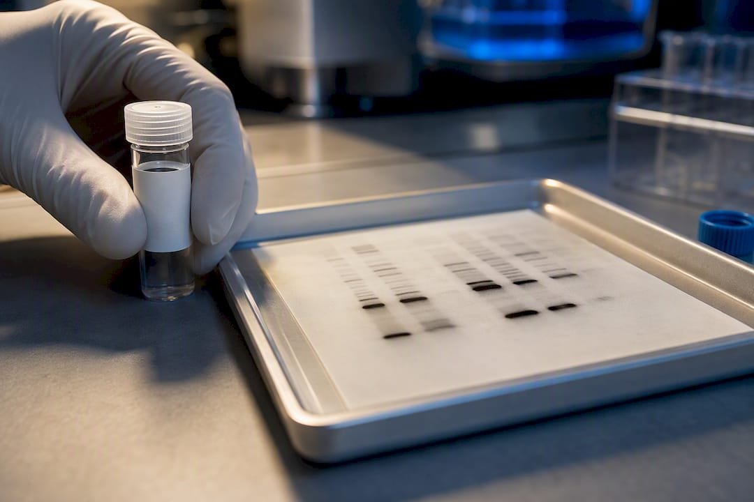
5 Validation Checks for Phospho-Specific Antibodies in the Lab
Phospho-specific antibodies recognize a target protein only when a defined serine, threonine, or tyrosine residue carries a phosphate group, making them the workhorse tool for tracking kinase and phosphatase activity in western blot, IHC, ELISA, and flow cytometry. They belong in your protocol whenever you need to know if a signaling event happened, not just whether a protein is present. The catch: without genetic controls, phosphatase treatment, or peptide competition, a clean band on a blot can still be lying to you.
TL;DR:
- Phospho-specific antibodies require proper validation, including peptide competition and genetic or enzymatic controls, to confirm true site-specific binding.
- Sample preparation must include phosphatase inhibitors and cold conditions to preserve phosphorylation states before analysis.
- Different assay formats like western blot, IHC, ELISA, and flow cytometry each have unique considerations, such as blocking reagents and fixation methods, affecting phospho-epitope detection.
- Lot-to-lot variability, especially in polyclonal antibodies, necessitates re-validation with controls for consistent and reliable results over time.
- Quantification should normalise phospho-signal to total protein levels and account for detector linearity to avoid misleading fold-change interpretations.
Table of Contents
- What Are Phospho-Specific Antibodies and How Are They Made?
- Why Use Phospho-Specific Antibodies in Your Experiments?
- How Do You Validate a Phospho-Specific Antibody?
- Sample Preparation: How Do You Preserve Phosphorylation Before It’s Gone?
- Assay-Specific Tips: Western Blot, IHC/IF, ELISA, and Flow
- Troubleshooting: Why Isn’t My Phospho-Antibody Working?
- How Should You Quantify Phospho-Antibody Signal?
- How Long Do Phospho-Specific Antibodies Stay Reliable in Storage?
- Why Do Antibody Lots Vary, and How Do You Manage It?
- A Lab Scientist’s Take on Chasing Antibody Specs
- Get Validated Antibodies and Peptide Controls From Mayflowerbio
- Sources
- FAQ
What Are Phospho-Specific Antibodies and How Are They Made?
A phospho-specific antibody is raised against a synthetic phosphopeptide, usually 10 to 15 residues long, centered on the modified serine, threonine, or tyrosine with the natural flanking sequence intact. Sequence context matters enormously here. The same phosphorylated residue in a different structural neighborhood can fold differently, so immunogen design has to account for the exact local sequence, not just the modified amino acid.
Production follows a few established paths, each with tradeoffs:
- Polyclonal antibodies are faster and cheaper to generate but vary batch to batch, since they represent a mixed population of B-cell responses.
- Monoclonal antibodies offer a single, reproducible epitope-binding clone, which matters for long-term studies spanning multiple antibody lots.
- Recombinant antibodies lock in the sequence at the DNA level, removing hybridoma drift entirely.
After generation, specificity depends on affinity purification: the antiserum gets run over a column bearing the phosphopeptide, then subtracted against the non-phosphorylated version of the same peptide to strip out antibodies that bind regardless of phosphorylation state. Current Protocols lays out this phosphopeptide immunogen design and purification workflow in detail, and it’s worth reading before you commission a custom antibody rather than after.
Why Use Phospho-Specific Antibodies in Your Experiments?
A positive signal from a validated phospho-specific antibody tells you a kinase or phosphatase acted on that exact residue at the moment you fixed or lysed your sample. That’s a fundamentally different piece of information than total protein abundance, and it’s why these reagents dominate signaling research.
Common applications, by format:
- Western blot captures activation kinetics: a time-course after growth factor stimulation, for instance, showing when a MAPK pathway member gets phosphorylated and when it resolves.
- IHC and IF map which cells, and which subcellular compartments, carry the phosphorylation event, which matters in heterogeneous tumor tissue.
- ELISA and multiplex platforms quantify phosphorylation across large cohorts, often as a pharmacodynamic marker showing whether a kinase inhibitor is hitting its target in patient samples.
- Mass spectrometry serves as the orthogonal check, confirming site-specific phosphorylation independent of any antibody, though it demands more sample and instrument time than antibody-based methods.
How Do You Validate a Phospho-Specific Antibody?
No single test proves specificity. Reviewers increasingly expect a combination of orthogonal approaches, and journals have started rejecting phospho-antibody data that relies on the antibody’s datasheet alone.
- Genetic controls first. A knockout or knockdown of the upstream kinase, or a phospho-dead mutant (serine or threonine swapped for alanine) at the target site, is treated as the gold standard because it removes the modification at the source rather than after the fact, a point reviewed extensively for western blot practice.
- Phosphatase treatment. Split your lysate, treat one half with a broad-spectrum phosphatase like lambda protein phosphatase, and confirm the signal disappears in the treated sample while total protein stays flat.
- Peptide competition. Pre-incubate the antibody with excess phosphopeptide (it should block signal) and with the non-phosphorylated equivalent (it should not) to demonstrate the antibody discriminates by phosphorylation state, not sequence alone.
- Orthogonal confirmation. Where feasible, confirm the same site by targeted mass spectrometry or a second antibody raised against an independent epitope on the same phosphoprotein.
- Document it. State in your methods exactly which controls you ran, lot numbers used, and any lot-to-lot re-validation, since combining multiple complementary validation strategies is what separates defensible data from a pretty blot.
Pro Tip: Ask your supplier for their validation dataset before you buy, not after a failed experiment. If they can’t produce phosphatase-treated or knockout-validated data, treat their datasheet claims as a starting hypothesis, not a fact.
Sample Preparation: How Do You Preserve Phosphorylation Before It’s Gone?
Phosphorylation is transient by biological design, which means your sample prep window is often the single biggest variable in whether your antibody works at all. The most frequent reason phospho-detection fails isn’t a bad antibody. It’s a lysate that lost its phosphorylation before the antibody ever touched it.
Practical steps that matter:
- Add broad-spectrum phosphatase inhibitor cocktails to lysis buffer immediately, not after you’ve finished harvesting all your plates.
- Keep everything on ice or at 4°C from harvest through lysis; endogenous phosphatases stay active at room temperature.
- Build lysis buffer around a detergent (RIPA or NP-40 based, depending on whether you need nuclear fractions), physiological salt, and a combined protease/phosphatase inhibitor mix.
- Snap-freeze in liquid nitrogen if you can’t process immediately, and never subject the same aliquot to repeated freeze-thaw cycles.
- Switch from PBS to TBS when probing phosphoepitopes; phosphate-buffered saline can interfere with phospho-specific detection, a caution documented in western blot validation literature.
If you need a positive control and don’t have access to a reliable stimulation paradigm, an in vitro approach using ATP addition to shift the kinase/phosphatase equilibrium in lysates can generate a phosphorylation-positive sample without live-cell stimulation, and it works across WB, ELISA, and IF.
Assay-Specific Tips: Western Blot, IHC/IF, ELISA, and Flow
Each platform has its own failure points, and generic antibody advice tends to skip over them.
For western blot, skip nonfat dry milk as a blocking reagent. Milk contains casein, which can cross-react with some phospho-specific antibodies and increase background signal. Use a phosphoprotein-free blocker such as BSA instead, and pair with a matched secondary antibody at the manufacturer’s recommended dilution.
For IHC and IF, fixation timing and antigen retrieval method both affect whether the phosphoepitope survives intact. Phosphoepitopes are notably labile, and pathology-focused reviews of phospho-specific antibody use flag this as a recurring source of false negatives in archived tissue. Tyramide-based signal amplification can help recover weak signal in low-abundance targets; Mayflowerbio’s tyramide reagent options are worth reviewing if your target sits near the detection floor.
For ELISA and multiplex, run calibration standards alongside a dephosphorylated negative control in every plate, and check cross-reactivity against related phosphoproteins in your panel.
For flow cytometry, use a fixation and permeabilization chemistry validated for phosphoepitopes; standard methanol-based protocols can strip some phospho-signals that milder paraformaldehyde-based kits preserve.
Pro Tip: If a phospho-blot looks unusually clean with almost no background, be more suspicious, not less. Overly clean phospho-blots sometimes mean the antibody isn’t binding the target at all.
Troubleshooting: Why Isn’t My Phospho-Antibody Working?
Work through failures in this order before concluding the antibody itself is the problem.
- Weak or absent signal: check whether phosphatase inhibitors were added at harvest and whether the sample sat at room temperature too long before lysis.
- High background: switch blocking reagent away from milk-based blockers and confirm you’re using TBS rather than PBS for the wash and block steps.
- Unexpected bands at the wrong molecular weight: run peptide competition alongside a genetic control; if the band persists after competition with excess phosphopeptide, it’s likely off-target binding, not your protein of interest.
- Inconsistent results across experiments: request certificate-of-analysis data for the specific lot you’re using and re-run your phosphatase-treatment control on any new lot before trusting comparative data.
How Should You Quantify Phospho-Antibody Signal?
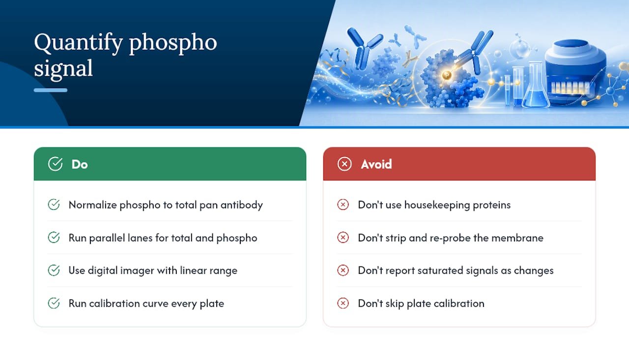
Raw band intensity from a phospho-western tells you almost nothing on its own, because it conflates two variables: how much total protein you loaded and how much of that protein is phosphorylated. The fix, recommended in guidance from the Journal of Biological Chemistry, is to normalize phospho-signal to a total protein measurement using a pan antibody against the same target, rather than relying on housekeeping proteins like actin or tubulin.
Stripping and re-probing the same membrane for total protein after phospho-detection is common practice, but it’s not the most reliable route. Stripping efficiency varies membrane to membrane, and residual phospho-signal can bleed into your total-protein readout and skew the ratio. Running parallel lanes, one probed for phospho-signal and one for total protein from the same lysate, avoids that artifact entirely, though it costs you gel real estate.
Dynamic range is the other quiet failure point. Phospho-signals can saturate on film or on some chemiluminescence imagers well before total protein does, especially with a highly abundant, highly modified target like phospho-ERK after strong stimulation. A digital imager with a documented linear range, and a dilution series run alongside your samples to confirm you’re inside that range, prevents you from reporting fold-changes that are really just detector saturation.
For multiplex or ELISA-based quantification, a calibration curve built from a recombinant phosphoprotein standard, run on every plate rather than once per antibody lot, catches plate-to-plate drift before it contaminates a cohort-wide comparison. None of this is exotic. It’s the difference between a phospho-blot that looks convincing and one that would survive a reviewer asking for the total-protein normalization data.
How Long Do Phospho-Specific Antibodies Stay Reliable in Storage?
Most phospho-specific antibodies ship as glycerol stocks meant for storage at negative 20 degrees Celsius, and the biggest threat to long-term performance isn’t age. It’s how many times you’ve frozen and thawed the vial.
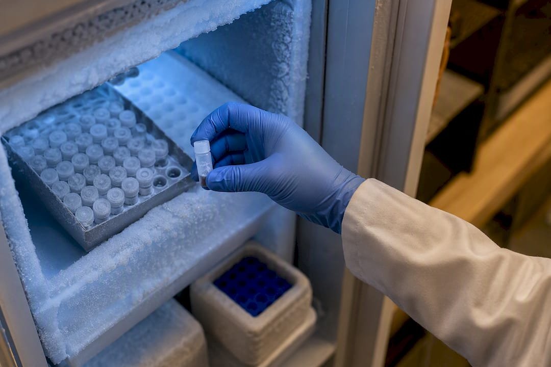
Repeated freeze-thaw cycles degrade antibody structure gradually, and for a reagent that already depends on recognizing a subtle conformational feature (a phosphorylated residue in its native context), that degradation shows up faster than it would for a antibody recognizing a simple linear epitope. Aliquoting a new vial into single-use volumes the day it arrives, rather than repeatedly dipping into one working stock, is the single easiest thing a lab can do to extend usable antibody life.
A few storage habits worth locking into your lab’s SOPs:
- Keep working aliquots at 4°C only for short-term use (days to a couple of weeks), and return to negative 20 for anything longer.
- Avoid storing antibody dilutions in the same buffer you’ll block with; some blocking proteins can encourage aggregation over time.
- Check for visible precipitate before use. It’s not always a dealbreaker, but it’s a signal to re-validate the vial’s activity with a positive control before trusting new results.
- Track opening date and number of freeze-thaw cycles on the vial label itself, not just in a digital log that nobody checks mid-experiment.
Light exposure matters less for most phospho-antibodies than it does for fluorophore-conjugated reagents, but conjugated versions (used in direct-labeled flow panels, for instance) do need protection from prolonged light exposure during both storage and bench use.
Why Do Antibody Lots Vary, and How Do You Manage It?
Lot-to-lot variability is one of the least discussed problems in phospho-antibody work, and it’s also one of the most common explanations for a result that worked beautifully in one experiment and fell apart in the repeat six months later, using a reagent from a different vial.
Polyclonal antibodies carry the highest lot variability by nature. Each new immunization produces a genuinely different mixture of antibody clones, even from the same animal, so a new lot can have different affinity, different background characteristics, or a subtly different specificity profile than the one it replaced. Monoclonal and recombinant antibodies reduce this risk substantially, since the clone itself doesn’t change, but manufacturing conditions (expression system batch, purification run) can still introduce small shifts in activity or aggregation state.
The practical fix is treating every new lot as if it were a new reagent, not a refill. Run your phosphatase-treatment control and, if you have one, your peptide-competition control on the new lot before using it for anything you plan to publish. Compare signal intensity against a banked sample from the previous lot, if you kept one, rather than assuming continuity. Request a certificate of analysis or lot-specific validation data from your supplier; a reputable vendor should be able to tell you which validation assays that specific lot passed, not just what the antibody is generally expected to detect.
For studies spanning years or multiple cohorts, some labs bulk-purchase a single large lot up front specifically to avoid this problem, accepting the storage burden in exchange for reagent consistency across the entire dataset.
A Lab Scientist’s Take on Chasing Antibody Specs
Too many labs chase a higher-affinity antibody when their real problem is upstream: a lysate that sat at room temperature for twenty minutes before anyone thought about phosphatase inhibitors. No antibody, however well validated, rescues a sample that already lost its phosphorylation before you ever added a reagent to it.
If you take one thing from this piece, take the validation-first mindset. A phospho-antibody with modest affinity but a documented phosphatase-treatment control and a clean peptide-competition result will give you more defensible data than a “premium” antibody with no controls behind it at all. Mayflowerbio’s approach reflects that priority: we’d rather point a researcher toward the right control peptide than sell a reagent without the tools to prove it worked. If you’re setting up a validation series, start a peptide competition control before you commit a full cohort’s samples to a single antibody lot.
— Alina
Get Validated Antibodies and Peptide Controls From Mayflowerbio
Mayflowerbio supplies the antibodies, peptides, and assay reagents that make the validation checklist above practical instead of theoretical, not just the antibody on its own.

Our antibody catalog covers targets across signaling, infectious disease, and cell biology research, and our technical team can help identify the right non-phosphorylated peptide counterpart for competition assays before you commit a full experiment to a single lot. If you’re building out a signaling panel, our multiplex assay resources cover how to design a phospho-readout panel without cross-reactivity headaches, and our inhibitors and modulators line stocks the phosphatase and kinase inhibitors your lysis buffer needs. Reach out through our contact form for validation guidance or a quote on custom peptide controls; it costs nothing to ask before you order.
Sources
- Antibody validation for Western blot: By the user, for the user
- Generation and Utilization of Phosphorylation State–Specific Antibodies to Investigate Signaling Pathways
FAQ
What are phospho-specific antibodies?
They’re antibodies that bind a target protein only when a specific serine, threonine, or tyrosine residue is phosphorylated, letting researchers detect an active signaling event rather than just protein presence.
How do you validate a phospho-specific antibody for publication?
Combine genetic knockout or knockdown controls, phosphatase treatment, and peptide competition, then confirm with an orthogonal method like mass spectrometry or an independent-epitope antibody where feasible.
What does it mean if a protein is phosphorylated?
A phosphate group has been added to a serine, threonine, or tyrosine residue, usually by a kinase, and that modification often switches the protein’s activity, localization, or interactions on or off.
Why does my phospho-antibody give high background on western blot?
Nonfat dry milk contains casein, a phosphoprotein that can cross-react with phospho-specific antibodies; switching to a BSA-based blocker and TBS wash buffer usually resolves it.
Can I generate a positive control without stimulating live cells?
Yes. Adding ATP to a lysate to shift the kinase/phosphatase equilibrium can produce a phosphorylation-positive sample suitable for antibody screening across several assay formats.




