
Antigen vs Antibody Tests: A Lab Selection Guide
For time-sensitive detection of a target pathogen or protein, use antigen-detection formats: sandwich ELISA, chemiluminescent immunoassays, or an optimized lateral flow immunoassay (LFIA). For measuring host immune response or prior exposure, use antibody-detection (serology) formats: indirect ELISA, TR-FRET, or a validated chemiluminescent platform.
The choice hinges on what molecule you’re actually measuring and when, relative to exposure. Antigen tests detect the pathogen or protein itself, so they work best on fresh, acute-phase samples. Antibody tests detect the host’s immune response, which makes them the right tool for retrospective or convalescent-phase analysis.
- Antigen formats: sandwich ELISA, chemiluminescent immunoassay, optimized LFIA
- Antibody formats: indirect ELISA, TR-FRET, validated chemiluminescent serology
- Sensitivity driver: antibody binding affinity, not concentration
- Supplier signal: Mayflowerbio stocks matched antibody pairs and antigen reagents suited to both formats
Key Takeaways
Antibody binding affinity, not antibody concentration, is the primary factor that determines whether an immunoassay hits its target sensitivity.
| Point | Details |
|---|---|
| Match analyte to format | Use antigen detection for acute, time-sensitive measurement and antibody detection for exposure history. |
| Affinity beats concentration | Raising a low-affinity antibody’s concentration usually increases background instead of fixing sensitivity. |
| Sandwich needs matched pairs | Sandwich ELISA gives the highest sensitivity but requires validated, non-overlapping capture/detection antibodies. |
| Preserve antigen conformation | Antigen-down coating must avoid denaturation or sensitivity drops sharply in antibody-detection assays. |
| Source documented reagents | Mayflowerbio provides batch-specific QC data and matched antibody pairs for infectious disease assay development. |
Table of Contents
- Antigen vs Antibody Tests: Comparing Lab Dimensions That Matter
- What Assay Formats Should You Use for Each Test Type?
- How Do You Select Antibodies and Antigens for Assay Development?
- What Validation Steps and Controls Prevent Assay Failures?
- What Belongs on an RFP for Antigen and Antibody Test Reagents?
- Where to Source Antigen and Antibody Reagents for Your Lab
- Sources
- FAQ
Antigen vs Antibody Tests: Comparing Lab Dimensions That Matter
The two assay types diverge on nearly every practical axis a lab manager cares about: what gets measured, how fast you get an answer, and how much development work sits between you and a validated protocol.
Antigen assays measure a specific protein or pathogen component directly, which is why they suit acute-phase or environmental samples where the analyte is present at detectable concentration right now. Antibody assays measure the host’s own immunoglobulin response, so they’re the better fit for tracking exposure history, vaccine response, or convalescent-phase serology. Combining both data streams often sharpens interpretation: an antigen-positive, antibody-negative result flags early or acute-phase material, while antigen-negative, antibody-positive suggests resolved or past exposure.
- Antigen assays generally need less serum/plasma volume and less sample prep than paired serology panels.
- Antibody assays tolerate a wider post-exposure sampling window, which matters for retrospective cohort studies.
- Running both in parallel gives a cleaner picture of infection stage than either alone, particularly in longitudinal studies.
| Dimension | Antigen detection | Antibody detection |
|---|---|---|
| Analyte detected | Target protein/pathogen | Host immunoglobulins (IgM/IgG) |
| Optimal sample timing | Acute phase, near time of exposure | Days to weeks post-exposure onward |
| Sensitivity/LOD pattern | High with sandwich formats; affinity-dependent | High with TR-FRET or chemiluminescence |
| Readout | Often quantitative (chemiluminescent, ELISA) | Often quantitative or titer-based |
| Throughput | Fast; LFIA gives near-immediate results | Moderate; plate-based formats take hours |
| Development complexity | Requires matched capture/detection pairs | Requires purified antigen, conformational integrity |
| Cost/supply-chain | LFIA components are cheap; matched pairs can be scarce | Antigen purity and lot consistency drive cost |
What Assay Formats Should You Use for Each Test Type?
Each format trades speed, sensitivity, and development effort differently, and picking the wrong one is the single most common reason assay development stalls.
Sandwich ELISA captures the analyte between two antibodies targeting different epitopes, then detects the bound complex with an enzyme-linked reporter. It’s the workhorse for antigen quantification because it typically delivers the highest sensitivity of any ELISA variant, but it demands a validated matched antibody pair, which is often the bottleneck in early development.
Indirect ELISA is the standard antibody-detection format: you coat a plate with antigen, then let patient or research serum bind, followed by a labeled secondary antibody. Direct ELISA skips the secondary step, trading some signal amplification for simpler protocols. Competitive ELISA works well when matched pairs aren’t available or when the analyte is a small hapten too small to support two simultaneous antibody binding sites; the standard curve slopes downward, and it generally uses less antibody overall.
LFIA (lateral flow immunoassay) delivers a result in minutes using capillary flow across a nitrocellulose membrane, but it trades quantitative precision for speed and needs careful reporter and membrane optimization to avoid false negatives. Newer nanobody-based rapid antigen platforms can hit correlation with ELISA results in under 30 minutes, though they still give up some sensitivity compared with a fully optimized plate assay.
Chemiluminescent immunoassays swap enzymatic color development for light-emitting substrates, pushing detection limits lower than standard colorimetric ELISA. TR-FRET (time-resolved fluorescence resonance energy transfer) is a homogeneous, no-wash antibody-detection format that can deliver results in about 15 minutes while suppressing background fluorescence, making it a strong candidate when throughput matters more than a lengthy validated workflow.
- Match the format to your matrix: serum and plasma behave differently on nitrocellulose than on polystyrene plates.
- Reporter choice (HRP/TMB vs. fluorophore vs. nanoparticle) should track your available detection instrument, not the other way around.
Pro Tip: A plate-based proof of concept rarely transfers directly to a lateral flow format. Membrane chemistry and reporter molecules almost always need re-optimization, or you’ll see false negatives that have nothing to do with the antibody’s actual performance.
How Do You Select Antibodies and Antigens for Assay Development?
Reagent quality determines whether your assay hits target sensitivity on the first attempt or burns three months in optimization. Start with antibody affinity, not price or brand recognition.
Affinity, expressed as the dissociation constant (KD) along with on/off binding kinetics, is the single strongest lever on assay sensitivity. Clonality matters too: monoclonal antibodies give you epitope specificity and lot consistency, while polyclonals can offer broader epitope coverage useful in some capture applications. For any sandwich format, demand documented evidence of a matched pair, meaning the capture and detection antibodies bind non-overlapping epitopes without steric interference.
Antigen source and purity matter just as much. Recombinant antigens with clear tag information (His, GST, Fc-fusion) give you predictable behavior in coating and conjugation steps, and purity data should accompany every lot. For antigen-down assays used in antibody detection, coating conditions must preserve the antigen’s native conformational epitopes; passive adsorption that denatures the protein can crater sensitivity before you’ve run a single sample.
Reporter and substrate choice should track your target limit of detection and available instrumentation. HRP with TMB substrate remains standard for colorimetric ELISA. Chemiluminescent substrates and fluorophores push detection lower for chemiluminescent platforms, and nanoparticle labels (quantum dots, lanthanide-doped particles) are expanding what LFIA can do quantitatively.
Before placing an order, request:
- Batch-specific QC data and a certificate of analysis for the exact lot you’ll receive
- Documented lot-to-lot consistency data, ideally with spike-recovery figures
- Matrix-matched validation data (serum, plasma, or your specific sample type)
- Recommended storage and handling conditions with stability data
- Epitope mapping or matched-pair confirmation for sandwich applications
Mayflowerbio’s infectious disease antibody listings include this documentation upfront, which shortens the reagent evaluation cycle considerably.
Pro Tip: Raising the concentration of a low-affinity antibody almost never fixes a weak signal. It usually just raises background and non-specific binding, since affinity and kinetics, not sheer quantity, drive sensitivity.
What Validation Steps and Controls Prevent Assay Failures?
A validation protocol that skips matrix-matched calibrators is a validation protocol waiting to fail in the field. Build these elements into every assay before you trust its output:
- Standards and calibrators spanning your expected dynamic range, ideally matrix-matched to the actual sample type
- Positive and negative controls run on every plate, not just during initial validation
- Spike-and-recovery experiments to catch matrix interference early
- Linearity checks across dilutions and intra/inter-assay coefficients of variation to confirm reproducibility
- A defined limit-of-detection estimate based on blank replicates, not a vendor’s generic spec sheet
Protocol-level details matter as much as the reagents themselves. Blocking strategy determines how much non-specific binding shows up as false signal, wash step optimization prevents carryover between wells, and plate coating conditions directly affect whether antigen-down formats retain conformational integrity.
Sandwich ELISA typically delivers higher sensitivity than competitive formats, a pattern consistent enough that it’s a reasonable default assumption during format selection, though competitive assays remain the better fit for small haptens.
Common failure modes and their usual fixes:
- High background: often traces back to insufficient blocking or a low-affinity antibody run at excessive concentration.
- Low signal: check antigen coating integrity first; denatured conformational epitopes are a frequent, overlooked culprit.
- Hook/prozone effects: high analyte concentrations can paradoxically suppress signal in sandwich formats; dilute and re-run before concluding the sample is negative.
- Inconsistent LFIA test lines: usually points to membrane lot variability or reporter conjugation inconsistency.
- Lot-to-lot reagent drift: request bridging samples from your supplier before switching lots mid-study.
What Belongs on an RFP for Antigen and Antibody Test Reagents?
A tight purchase order saves weeks of back-and-forth with a supplier’s technical team. Specify these details every time:
- Reagent format (antigen, monoclonal or polyclonal antibody, conjugate) and host species
- Conjugation state and label type (HRP, biotin, fluorophore)
- Lot size and available storage/shipping conditions (cold chain requirements)
- Required QC metrics: purity percentage, endotoxin levels, activity data
- Technical support SLA and expected lead time
Before finalizing a vendor, ask directly whether they offer matched antibody pairs with documented epitope mapping, whether they can supply lot-bridging samples when you need to switch batches mid-study, and whether their recommended protocol has been validated in your specific sample matrix.
- Confirm alternate supply options exist in case of a stock-out; single-source dependency on a niche antibody clone is a real project risk.
- Ask about typical lead times for custom conjugation or bulk lot reservations.
A Supplier’s Perspective on Antigen and Antibody Assay Support
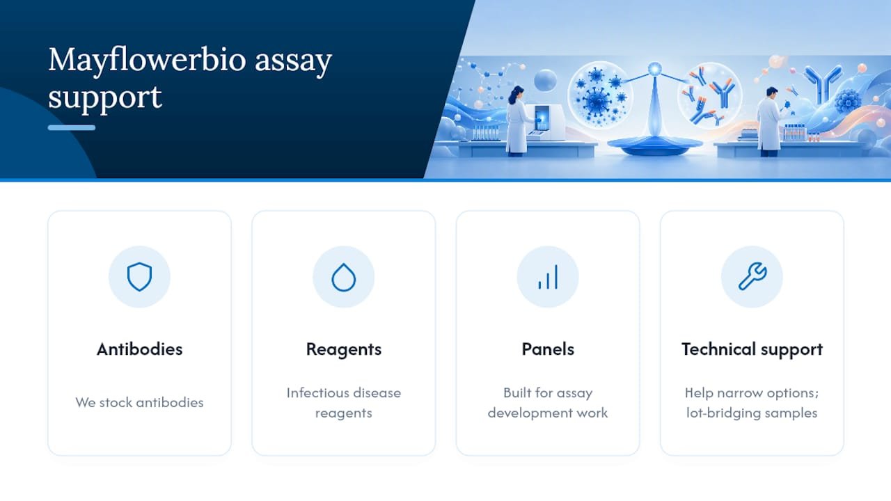
At Mayflowerbio, we stock antibodies, infectious disease reagents, and multiplex bioassay panels built for exactly this kind of assay development work. If you’re troubleshooting a matched-pair search or need lot-bridging samples for a study already underway, our technical support team can help you narrow the options.
Where to Source Antigen and Antibody Reagents for Your Lab
Choosing the right test format only gets you halfway. The reagent itself, and the documentation behind it, determines whether your assay hits its sensitivity target on the first run or needs three rounds of troubleshooting.

Mayflowerbio’s antibody catalog covers infectious disease targets with batch-specific QC data attached to every listing, so you’re not chasing a certificate of analysis after the order ships. For labs building multiplex panels that combine antigen and antibody readouts in one run, the bioassays category includes AimPlex multiplex kits designed for exactly that use case. If you need a matched pair for a sandwich ELISA or a lot-bridging sample to validate against your current reagent, reach out to our technical support team directly, they can walk you through availability and recommended protocols before you commit to a purchase order.
Sources
- Immunoassay Methods – Assay Guidance Manual – NCBI Bookshelf
- Lateral Flow Immunoassays for Detecting Viral Infectious Antigens and Antibodies – PMC
- Development of a broadly adaptable TR‑FRET serological assay – Sensors & Diagnostics (RSC)
This article is general information, not a substitute for advice from a qualified doctor. Consult a qualified healthcare professional about your own circumstances before acting on anything here.
FAQ
What Is the Main Difference Between Antigen and Antibody Tests?
Antigen tests detect the target pathogen or protein directly, while antibody tests detect the host’s immune response to that pathogen, making antigen tests better for acute-phase detection and antibody tests better for exposure history.
How Are Antigen Tests Done in a Research Lab?
Antigen tests typically use sandwich ELISA, chemiluminescent immunoassay, or LFIA, capturing the target protein between matched antibodies or across a lateral flow membrane for a rapid readout.
What Determines Antigen Test Accuracy?
Antibody binding affinity and the quality of the matched capture/detection pair drive sensitivity far more than antibody concentration or substrate choice alone.
How Do You Interpret Antibody Test Results Alongside Antigen Data?
An antigen-positive, antibody-negative pattern typically indicates early or acute-phase material, while antigen-negative, antibody-positive results suggest resolved or past exposure, so running both in parallel sharpens interpretation.
Where Can Labs Source Matched Antibody Pairs and Antigens?
Mayflowerbio supplies antibodies and infectious disease reagents with batch-specific QC documentation, and its technical support team can help identify matched pairs or lot-bridging samples for ongoing studies.




