
Cell Counting Dyes for Labs: Erythrosin B and the 3–5 Minute Rule
Trypan blue remains the default for manual and bright-field automated counting, with erythrosin B as the safer alternative when toxicity or automation optics are a concern, and fluorescent dyes like propidium iodide (PI) or 7-AAD, which are used for flow-based work.
TL;DR:
- Trypan blue remains the standard dye for manual and bright-field automated cell counting, but erythrosin B offers similar accuracy with lower toxicity.
- Accurate hemocytometer counts require mixing the dye thoroughly, loading immediately, and counting within 3 to 5 minutes to prevent dye uptake by live cells.
- Fluorescent dyes like propidium iodide and 7-AAD are used specifically for flow cytometry and multiplexing, not bright-field counting.
- Dye-exclusion assays alone do not reveal early apoptotic cells or metabolic activity, so combining with other viability tests improves reliability.
- Automated counters need calibration adjustments when switching dyes, and manual cross-checks help prevent inaccurate data from protocol changes.
Table of Contents
- What Are the Best Cell Counting Dyes for Different Applications?
- How Do You Count Cells With Trypan Blue on a Hemocytometer?
- When Should You Use Erythrosin B or Fluorescent Dyes Instead?
- Why Dye-Exclusion Assays Don’t Tell the Whole Viability Story
- Getting Reliable Results From Automated Cell Counters
- Safety and Disposal Notes for Cell Counting Dyes
- Mayflower Bioscience Resources for Reliable Cell Counting
- What Actually Matters When You Pick a Counting Dye
- Where to Find the Right Reagents and Slides for Your Workflow
- Sources
- FAQ
What Are the Best Cell Counting Dyes for Different Applications?
Not every dye fits every platform, and picking the wrong one wastes time or, worse, gives you numbers you can’t defend in a paper. Here’s how the common options stack up.
- Trypan blue stains dead cells whose membranes have lost integrity, letting them absorb the dye while live cells stay clear. It’s the workhorse for hemocytometers and most bright-field automated counters, but it carries documented health and environmental hazards that have pushed labs to look for substitutes.
- Erythrosin B works on the same exclusion principle as trypan blue but shows comparable counting accuracy with lower cytotoxicity, making it a reasonable swap for routine QC counts.
- Propidium iodide (PI) is a fluorescent nucleic acid stain that only enters cells with compromised membranes. It’s a staple for flow cytometry and fluorescence-based automated counters, not bright-field systems.
- 7-AAD behaves similarly to PI but with a different excitation/emission profile, useful when you’re multiplexing with other fluorophores in a flow panel.
- Eosin is mostly a histology counterstain rather than a viability dye, though some labs use it for quick bright-field contrast in tissue prep.
- Methylene blue sees occasional use in microbiology and older cytology protocols, but it’s not standard for mammalian cell viability counting today.
If you’re running a manual hemocytometer count for routine passage checks, trypan blue is still the fastest path. If you’re feeding cells into an automated imager all day, erythrosin B cuts your exposure risk. If you need single-cell resolution alongside surface markers, PI or 7-AAD on a flow cytometer is the only real option.
How Do You Count Cells With Trypan Blue on a Hemocytometer?
The standard protocol is short, but precision in timing and mixing matters more than most people assume. Here’s the sequence:
- Prepare your cell suspension and bring 0.4% trypan blue to room temperature.
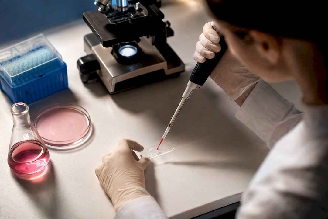
- Mix cells and dye at a 1:1 ratio (commonly 10 µL cell suspension plus 10 µL dye).
- Load 10 µL of the mixture into a clean hemocytometer chamber immediately after mixing.
- Count within 3 to 5 minutes. Waiting longer lets live cells start absorbing dye, inflating your dead-cell count.
- Count cells in all four corner squares (16 small squares each) under 10x magnification, applying a consistent rule for cells touching the boundary line.
- Average the counts across the four corner squares.
- Calculate cells per mL: average count × 10,000 × dilution factor.
A worked example: if your average corner-square count is 45 cells and you diluted 1:2, that’s 45 × 10,000 × 2 = 900,000 cells/mL. Simple arithmetic, but it only holds up if your mixing was even and you avoided bubbles when loading the chamber. A cell counting slide with defined chamber depth removes some of the loading variability that trips up manual counts.
When Should You Use Erythrosin B or Fluorescent Dyes Instead?
Erythrosin B earns its place when trypan blue’s toxicity profile or its interference with automated optics becomes a problem. Published comparisons found EB produces counting accuracy on par with trypan blue while showing lower cytotoxicity in cultured cells, and imaging-based workflows report that concentrations as low as 0.005% to 0.015% w/v work well with automated imagers once exposure and thresholding are tuned.
- Classic manual protocols use EB in the 0.1% to 0.4% range, similar to trypan blue.
- Automated imaging pipelines often need far lower concentrations, down to 0.005% w/v, for accurate linearity.
- PI and 7-AAD make sense when you need flow cytometry compatibility or plan to multiplex with fluorescence assays, but check spectral overlap before adding a third or fourth fluorophore to your panel.
Why Dye-Exclusion Assays Don’t Tell the Whole Viability Story
Dye-exclusion measures one thing: membrane integrity. It doesn’t capture metabolic activity, and that gap matters more than most protocols admit. A cell in early apoptosis can still exclude trypan blue or erythrosin B even though it’s already committed to dying, which means your “live” count can quietly overstate actual health.
- Pair dye-exclusion counts with a metabolic assay (resazurin or ATP-based) or Annexin V/flow cytometry when the result feeds into a publication.
- Run at least two to three technical replicates per sample rather than trusting a single count.
- Keep your counting window consistent across every sample in a batch. A count taken at 2 minutes and another at 6 minutes are not comparable data.
The literature on cell viability assays is consistent on this point: exclusion assays are a fast screen, not a final verdict.
Pro Tip: Run a pilot comparison between trypan blue and your chosen alternative on each new cell line before switching protocols. Some lines take up dye differently, and a swap that worked fine for HEK293 cells can throw off counts on a primary culture.
Getting Reliable Results From Automated Cell Counters
Most bright-field automated counters are built and calibrated around trypan blue detection, so switching dyes without adjusting instrument settings is a common source of bad data.
- Confirm your counter’s software thresholds match the dye you’re using. Erythrosin B often needs different exposure and contrast settings than trypan blue.
- Run a known-viability QC sample at the start of each session to catch calibration drift early.
- Fluorescence-based counters need PI, 7-AAD, or an equivalent stain. Trypan blue and erythrosin B won’t register on a fluorescence channel.
- Fall back to manual hemocytometer counts periodically to cross-check automated results, especially after any protocol change.
Understanding why counting accuracy matters for downstream data quality makes it easier to justify the extra QC step to a lab manager who wants faster throughput.
Safety and Disposal Notes for Cell Counting Dyes
Trypan blue’s safety profile is worth taking seriously, not just filing away. Reported concerns include potential teratogenic effects, which is part of why erythrosin B has gained traction as a lower-risk substitute.
- Wear gloves and eye protection when handling any vital dye, and work under a fume hood if your institution requires it for hazardous stains.
- Dispose of dye-stained waste according to your institution’s hazardous-waste procedures, not down the sink.
- Biohazardous or GMO-derived stained material needs separate handling per your biosafety office’s rules.
- Contact your institutional EH&S office before switching dyes if you’re unsure how the new reagent fits your existing waste stream.
Mayflower Bioscience Resources for Reliable Cell Counting
Getting consistent counts depends as much on your consumables as your protocol. Mayflowerbio’s cell counting slides are built for consistent chamber depth, and the cell biology product line covers reagents that support both dye-exclusion and complementary viability workflows. Technical datasheets and protocol support are available through the site’s product pages for labs standardizing their counting methods.
What Actually Matters When You Pick a Counting Dye
Most labs overthink the dye choice and underthink the validation step. Trypan blue works fine for quick QC checks, but if a number is going into a manuscript, run a pilot comparison against erythrosin B or a metabolic assay on your specific cell line first. Reagent hazard sheets change; cell line behavior does too. Available reagent and slide lines can serve as a starting point for labs standardizing this workflow.
— Alina
Where to Find the Right Reagents and Slides for Your Workflow
Switching between trypan blue, erythrosin B, and fluorescent exclusion dyes shouldn’t mean juggling three different suppliers and hoping the datasheets line up.

Mayflowerbio’s cell biology category carries the reagents and counting slides referenced throughout this guide, backed by technical datasheets you can check against your own protocol before ordering. Labs standardizing a counting workflow across multiple cell lines can also review the protocols and QC resources available on the site to make sure new reagents fit existing calibration steps. If you’re weighing a switch to erythrosin B or adding a flow-based confirmatory assay, reach out through the product pages for technical support before you commit a full batch of samples to a new dye.
This article is general information, not a substitute for advice from a qualified doctor. Consult a qualified healthcare professional about your own circumstances before acting on anything here.
Sources
- Trypan blue dye exclusion assay: Protocols and best practices (PMC)
- Application of a non-hazardous vital dye for cell counting with automated cell counters (Analytical Biochemistry, 2016)
- Chapter on cell viability assays (NCBI Bookshelf)
- Image-Based High-Throughput Cell Viability Assay Using Erythrosine B (Synentec technical note)
FAQ
How much trypan blue should you use for cell counting?
Standard protocol calls for 0.4% (w/v) trypan blue mixed 1:1 with your cell suspension, such as 10 µL of dye to 10 µL of cells, counted within 3 to 5 minutes of mixing.
How accurate is manual hemocytometer cell counting?
Accuracy depends heavily on operator technique, mixing consistency, and counting within the recommended time window; results improve with technical replicates and cross-checks against an automated counter or metabolic assay.
Why do you multiply the hemocytometer count by 10,000?
The multiplication factor of 10,000 corrects for the fixed volume of the hemocytometer’s counting grid, which holds 0.1 microliters per square millimeter, converting your raw cell count into cells per milliliter.
Is trypan blue toxic?
Trypan blue carries reported health and environmental concerns, including potential teratogenic effects, which is why many labs use PPE, follow institutional disposal rules, and consider erythrosin B for routine automated counting.
Can erythrosin B replace trypan blue for all applications?
Erythrosin B works well as a substitute for bright-field manual and automated counting with comparable accuracy and lower cytotoxicity, but it doesn’t apply to flow cytometry work that requires fluorescent dyes like PI or 7-AAD.




