
Cytokine Stability: Practical Lab SOPs for Researchers
Cytokine stability is analyte-dependent and matrix-dependent, not universal, so the safest default is to separate plasma or serum, freeze it at −80°C in single-use aliquots, and get whole blood centrifuged within an hour of the draw. Skip that window and you risk measuring an artifact of handling rather than the biology you set out to study.
Four rules cover most of what goes wrong in practice:
- Process fast. Centrifuge within 60 minutes of collection; if you can’t, keep the tube at 2–8°C, not at room temperature.
- Aliquot once. Split every sample into single-use volumes at the time of freezing. Never refreeze a partially thawed tube.
- Default to −80°C. Reserve −20°C or 4°C for short, defined windows only, never as long-term storage.
- Run a sentinel. Include an internal control sample, processed identically to your experimental samples, on every plate and every storage batch.
Watch the outliers. IL-1β, IL-8, and several other analytes degrade measurably faster than the rest of a typical panel, so don’t assume your worst-behaved cytokine sets the safety margin for the whole set. A systematic review of pre-analytical cytokine handling found several cytokines starting to degrade within one to three years even at −80°C, which is why sentinel QC samples matter more than a calendar rule ever will.
Key Takeaways
Reliable cytokine measurement depends on fast processing, −80°C single-use storage, and analyte-specific handling, since no single rule protects every cytokine equally.
| Point | Details |
|---|---|
| Process within an hour | Centrifuge whole blood within 1 hour of the draw, or keep it cold and log the delay. |
| Default to −80°C | Long-term storage at −80°C outperforms −20°C and 4°C for nearly every cytokine tested. |
| Aliquot once, never refreeze | Single-use aliquots of 100 to 250 microliters prevent thaw-refreeze artifacts. |
| Watch labile analytes closely | IL-1β, IL-8, IL-13, and IL-17 degrade faster than the rest of a typical panel. |
| Use sentinel QC every batch | An internal control processed identically to experimental samples catches procedural failures early. |
| Validate archived samples before reuse | Spike-recovery and ICC/Bland-Altman comparisons confirm whether old specimens still reflect biology. |
| Build your SOP around Mayflowerbio resources | Mayflowerbio’s transport media, recombinant standards, and multiplex design guidance support each stage of a stability-focused workflow. |
Table of Contents
- Quick-Reference Table for Cytokine Storage Temperature and Duration
- What the Peer-Reviewed Literature Says About Cytokine Thermal Stability
- Why Cytokines Degrade: Mechanisms Behind Sample Instability
- How Should You Collect and Process Samples for Cytokine Analysis?
- What Storage Temperature Should You Use, and How Many Freeze-Thaw Cycles Are Safe?
- Which Cytokines Are Most Stable, and Which Degrade Fastest?
- How Does Assay Choice Interact With Storage History?
- What Should You Do When Cytokine Results Look Wrong?
- Copy-Ready SOP Checklist for Biobanking and Field Collection
- Where the Evidence Still Falls Short
- Mayflowerbio’s Recommended Default Settings for Cytokine Sample Handling
- How Mayflowerbio Supports Reliable Cytokine Measurement
- Sources
- FAQ
Quick-Reference Table for Cytokine Storage Temperature and Duration
Print this and tape it above the centrifuge. It won’t answer every question, but it covers the decisions that go wrong most often.
| Condition | Duration | Action |
|---|---|---|
| Whole blood, room temp | 0–1 hour | Acceptable; centrifuge promptly |
| Whole blood, room temp | >1 hour | Keep at 2–8°C, log the delay as an exception |
| Serum/plasma at 4°C | Hours to 1–2 days | Short-term only; measurable degradation begins by day 4 |
| Serum/plasma at −20°C | Weeks | Temporary bridge storage only, not a long-term solution |
| Serum/plasma at −80°C | Months to ~2 years | Preferred for archival and repeat testing |
| Liquid nitrogen (−150°C+) | Years | Reserve for irreplaceable biobank specimens |
A few things the table can’t show: aliquot at 100 to 250 microliters per tube so a single freeze-thaw cycle never touches more material than one assay run needs, and label every vial with draw date, processing time, and freezer location, not just a sample ID.
Pro Tip: Batch your aliquoting by cytokine panel, not by patient. If IL-1β and IL-17 are on your panel, plan for smaller, more numerous aliquots since those analytes tolerate fewer freeze-thaw cycles than more robust markers like IL-10.
What the Peer-Reviewed Literature Says About Cytokine Thermal Stability
The consensus across the literature is narrower than most SOPs assume: cytokine degradation is analyte-specific, and no single storage rule protects an entire multiplex panel equally. A review focused on multiplex immunoassay pre-analytics reported that most cytokines remain stable for extended periods at −80°C, but certain cytokines including IL-13, IL-15, IL-17, and CXCL8 may start degrading within one year under the same conditions. That gap between “most cytokines” and “the ones you actually care about” is where a lot of published data quietly loses its footing.
A prospective study of 24 donors examined eight cytokines and found significant degradation for several of them after just four days at 4°C, with IL-1β and IL-8 both dropping measurably after only two freeze-thaw cycles. The same study confirmed −80°C outperformed −20°C for every analyte tested. That’s a small sample size, and it’s worth treating the specific numbers as directional rather than definitive, but the direction is consistent with the broader literature.
Matrix effects complicate the picture further. Research comparing serum and plasma concentrations using ICC and Bland-Altman analyses found generally poor absolute agreement between the two matrices for many cytokines, and thawing itself significantly changed concentrations for most analytes tested. A few cytokines only showed acceptable agreement within narrow concentration ranges.
Cytokine stability doesn’t fail as a category. It fails one analyte at a time, in one matrix at a time, which is exactly why panel-wide assumptions keep tripping up otherwise careful studies.
Not every finding points toward fragility. A freeze-thaw study of 16 serum immunoregulators found six analytes, including CRP, IL-8, IL-10, IFN-γ, IP-10, and eotaxin-1, remained quantitatively equivalent across 25 and even 50 freeze-thaw cycles. That’s a genuinely useful finding for biobanks sitting on archived specimens they assumed were compromised.
Why do studies disagree? Three recurring culprits:
- Different matrices (serum versus EDTA plasma versus heparin plasma) behave differently for the same analyte.
- Different assay platforms (singleplex ELISA versus multiplex bead arrays) carry different cross-reactivity and sensitivity profiles.
- Donor-to-donor variability sometimes exceeds the variability introduced by storage conditions, as one multi-matrix Luminex study found when donor and matrix effects outweighed freeze-thaw effects across four cycles.
Why Cytokines Degrade: Mechanisms Behind Sample Instability
Degradation isn’t one process. It’s several competing ones, and understanding which is dominant in your samples tells you which SOP step actually matters most.
- Cellular release after the draw. Platelets and leukocytes continue releasing or consuming cytokines after blood leaves the vein, which is why delayed centrifugation artificially inflates some analytes and depletes others, according to research from the University of Minnesota’s cytokine lab.
- Proteolytic degradation. Serine proteases and other enzymes present in blood actively cleave certain cytokines over time, particularly in serum where clotting releases additional proteolytic activity.
- Receptor and protein binding. Some cytokines bind soluble receptors or carrier proteins, effectively hiding them from immunoassays without any true loss of the molecule itself, which muddies the line between “degraded” and “undetectable.”
- Adsorption to plastic. Certain cytokines stick to the walls of storage tubes, especially at low concentrations, silently reducing recoverable volume with every transfer.
- pH drift. Prolonged storage without proper sealing allows CO2 loss and pH shifts that can destabilize sensitive protein structures.
Matrix choice interacts with nearly all of these. Serum allows clotting-associated protease release that plasma avoids, while EDTA plasma chelates the divalent cations several proteases need to function, offering a modest protective effect. Heparin plasma sits in between, and citrate plasma introduces its own dilution and calcium-binding effects that complicate direct comparison to the other two.
Pro Tip: If you suspect matrix-driven artifacts, run a small spike-recovery experiment: add a known concentration of recombinant cytokine, like an IL-4 recombinant standard, into your serum and plasma matrices side by side, then measure recovery after your standard processing workflow. A recovery gap between matrices tells you more than any published paper will about your specific assay and specimen type.
How Should You Collect and Process Samples for Cytokine Analysis?
1. Choose the tube deliberately. EDTA plasma is the preferred matrix in many cytokine studies because EDTA chelates calcium and inhibits several proteases, reducing post-draw degradation compared to serum. Heparin plasma is an acceptable alternative but can interfere with certain multiplex platforms. Citrate tubes are typically reserved for coagulation studies, not cytokine panels, since the dilution factor complicates concentration calculations.
2. Hit the centrifuge window. Target centrifugation within one hour of the draw. Research on pre-analytical handling found that IL-17A and IL-6 concentrations shifted measurably after just three hours at room temperature, while keeping the same tubes at 2–8°C delayed or prevented much of that change. If you can’t hit the one-hour target, cold storage pending centrifugation is your fallback, not room temperature.
3. Centrifuge at defined, repeatable parameters. Most protocols call for 1,000 to 2,000 times gravity for 10 to 15 minutes at 4°C, though your specific tube manufacturer’s guidance should take precedence if it differs. Consistency across every batch matters more than chasing a theoretically optimal speed.
4. Aliquot immediately after spinning. Split the supernatant into single-use volumes right away rather than storing one large tube and drawing from it repeatedly. Every extra freeze-thaw cycle on a shared tube compounds risk across every assay run planned from it.
5. Record the metadata that actually gets used later. Time of draw, time to centrifuge, centrifuge speed and duration, aliquot volume, freezer ID, and freeze date and time. Missing metadata is the single most common reason archived cohorts become unusable years later.
6. Keep light exposure minimal. Extended exposure to ambient or direct light during processing can degrade light-sensitive proteins in a panel; process in normal lab lighting but avoid direct sunlight or prolonged bench exposure before freezing.
Pro Tip: When a delay is unavoidable, document it as a formal exception rather than silently absorbing it into your dataset. A flagged “processed at 90 minutes, kept cold” note lets you exclude or weight that sample later; an undocumented delay just becomes invisible noise in your results.
What Storage Temperature Should You Use, and How Many Freeze-Thaw Cycles Are Safe?
The temperature hierarchy is consistent across the literature: −80°C beats −20°C, which beats 4°C, for essentially every cytokine tested. Comparative work on 27 multiplex-assayed cytokines found storage at −20°C accelerated degradation relative to −80°C across repeated and long-term storage, with the gap widening the longer samples sat.
Liquid nitrogen storage (below −150°C) offers the longest theoretical stability window and is standard practice for biobanks holding irreplaceable cohort specimens for a decade or more, though the added cost and logistics rarely justify it for routine research storage where −80°C mechanical freezers perform well within a two-year window.
Freeze-thaw tolerance varies enormously by analyte, which is the detail most SOPs flatten into a single blanket rule. The freeze-thaw study of 16 serum immunoregulators mentioned earlier found six analytes held steady across 25 to 50 cycles, while others degraded well before that point. Meanwhile, an 80-plex Luminex study across multiple matrices found that donor variability and matrix choice contributed more variance than freeze-thaw itself for most cytokines across four cycles, with matrix-specific exceptions.
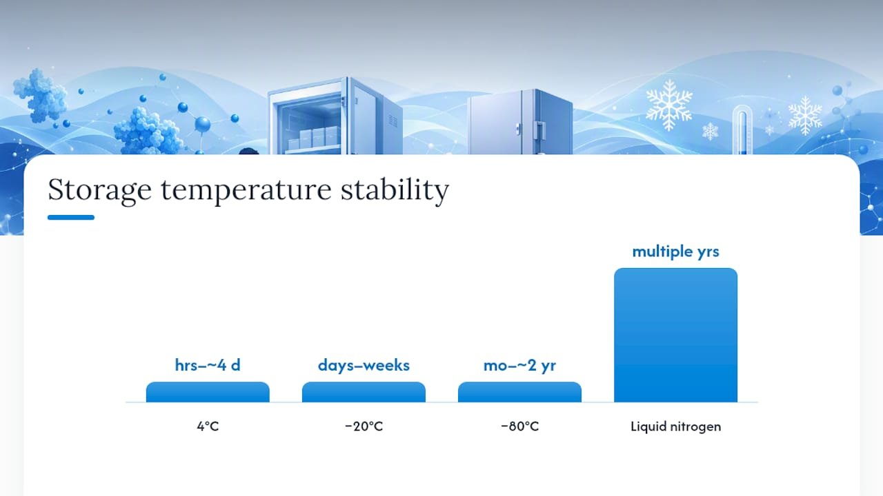
| Storage condition | Typical stability window | Freeze-thaw notes |
|---|---|---|
| 4°C | Hours to ~4 days | Not applicable; not a frozen state |
| −20°C | Days to weeks | Accelerated degradation versus −80°C on repeated cycling |
| −80°C | Months to ~2 years for most analytes | Most cytokines tolerate up to 4 cycles; sensitive analytes (IL-1β, IL-8) show loss after 2 |
| Liquid nitrogen | Multiple years | Best long-term option for irreplaceable biobank specimens |
Practical rules that follow from this:
- Treat any cytokine panel as having at least a few fragile members until proven otherwise in your own hands.
- Cap routine freeze-thaw cycles at two for panels containing IL-1β, IL-8, or other analytes with documented early degradation.
- Thaw on ice or at 4°C, never at room temperature or in a water bath, and move straight into assay setup once thawed.
- Never refreeze a partially used aliquot; that’s what single-use aliquoting exists to prevent.
The clearest single number to anchor your SOP around: cytokines held at −80°C remain broadly stable for roughly two years, but a handful of specific analytes start slipping within the first twelve months, according to the multiplex immunoassay prerequisites review. Build your retesting schedule around the fragile analytes, not the robust ones.
Which Cytokines Are Most Stable, and Which Degrade Fastest?
Not every analyte in a standard panel behaves the same way, and treating IL-6 and IL-17 as equally durable is one of the most common mistakes in archived-sample studies. Here’s a working rubric based on the available evidence:
- IL-1β: Labile. Degrades after as few as two freeze-thaw cycles and shows measurable loss within days at 4°C. Measure early, aliquot small.
- IL-4: Moderate stability. Behaves reasonably well at −80°C but shows matrix-dependent variability between serum and plasma.
- IL-5: Moderate. Generally holds up over short-term frozen storage but has less long-term data than more commonly studied analytes.
- IL-6: Variable, matrix-sensitive. Shifts have been documented within hours at room temperature before processing; keep pre-centrifuge time minimal.
- IL-7: Relatively understudied but generally grouped with moderate-stability interleukins in multiplex panels; validate locally.
- IL-8 (CXCL8): Labile. Drops after two freeze-thaw cycles and is one of the analytes flagged for degradation within a year even at −80°C.
- IL-10: One of the more robust analytes; showed equivalence across 25 to 50 freeze-thaw cycles in immunoregulator stability testing.
- IL-12p70: Heterodimeric structure makes it more susceptible to degradation than single-chain cytokines; handle conservatively.
- IL-13: Labile. Documented degradation within one year of −80°C storage in long-term stability data.
- IL-17: Labile and matrix-sensitive. Shows early degradation both in short delays before processing and in longer-term frozen storage.
- IFN-γ: Moderate to variable. Affected by processing delays beyond a few hours; agreement between serum and plasma is inconsistent across studies.
- TNF-α: Generally moderate stability at −80°C but shows serum-plasma discordance in comparative studies.
- MCP-1 (CCL2): Comparatively robust in most matrices, though data is less extensive than for the interleukins above.
- MIP-1β (CCL4): Moderate stability; matrix choice appears to matter more than freeze-thaw count for this chemokine.
- G-CSF: Generally stable under standard frozen storage, with fewer reports of rapid degradation than the labile interleukins above.
| Cytokine | Stability tendency | Matrix sensitivity | Handling note |
|---|---|---|---|
| IL-1β | Labile | High | Minimize freeze-thaw, aliquot small |
| IL-8 | Labile | Moderate | Cap at 2 freeze-thaw cycles |
| IL-10 | Robust | Low | Tolerates extensive freeze-thaw |
| IL-17 | Labile | High | Process quickly, avoid delay |
| IFN-γ | Variable | Moderate | Validate serum vs. plasma locally |
If your panel includes several of the labile analytes above, design your SOP around the weakest link, not the average.
How Does Assay Choice Interact With Storage History?
Multiplex bead arrays are efficient but more sensitive to matrix effects and cross-reactivity between analytes than singleplex ELISA, particularly once samples have been through multiple freeze-thaw cycles or extended storage. If a specific analyte in your panel is known to be marginal, running it separately by ELISA can sometimes recover cleaner data than leaving it bundled into a 20-plex panel where one degraded analyte’s signal drift can distort neighboring wells through cross-reactivity.
Before trusting archived samples with any assay format, run a short validation sequence:
- Spike-recovery testing. Add a known quantity of recombinant standard into a matrix-matched blank and confirm recovery falls within an acceptable range for your platform.
- Dilution linearity. Confirm serial dilutions of a high-concentration sample return proportionally lower readings; nonlinearity flags matrix interference.
- Comparison to fresh samples using ICC or Bland-Altman analysis. If you have any paired fresh versus archived samples, these statistical methods reveal systematic bias that a simple correlation coefficient can miss.
Reviewing your assay’s multiplex design principles before finalizing a panel helps catch cross-reactivity risks before they show up as unexplained batch effects months into a study.
Pro Tip: Run a sentinel sample, processed and stored identically to your experimental cohort, on every single plate. If your sentinel’s values drift outside your established range, you’ve caught an assay or storage problem before it contaminates your real data, not after.
What Should You Do When Cytokine Results Look Wrong?
1. Check the sentinel control first. If your batch-matched internal control falls outside its expected range, the problem is likely procedural, not biological, and results from that batch need review before interpretation.
2. Review the metadata for the flagged samples. Extended time-to-centrifuge, an undocumented storage gap, or an unusual number of freeze-thaw cycles often explains an outlier better than a biological hypothesis does.
3. Match the failure pattern to a likely cause. A uniformly elevated inflammatory marker across a batch often points to delayed processing and cellular release; a uniformly depressed result across older samples often points to storage-related degradation rather than a true biological signal.
4. Decide: re-assay or exclude. If archived material remains and the analyte is one of the more freeze-thaw-tolerant ones, a re-assay after confirming proper thaw procedure may be worthwhile. For labile analytes like IL-1β or IL-17 that have already gone through multiple cycles, exclusion is usually the more defensible choice.
Pro Tip: Keep a running log of which sample batches fail sentinel QC and why. Patterns across batches, not single failures, are what tell you whether the issue is a reagent lot, a freezer malfunction, or a genuine biological signal worth chasing.
Copy-Ready SOP Checklist for Biobanking and Field Collection
1. Draw blood into the appropriate anticoagulant tube for your panel (EDTA preferred for most cytokine work).
2. Centrifuge within 1 hour; if delayed, store at 2–8°C and log the exception.
3. Aliquot supernatant immediately into single-use volumes of 100 to 250 microliters.
4. Freeze at −80°C within 30 minutes of aliquoting.
5. Record metadata: draw time, centrifuge parameters, aliquot ID, freezer ID, freeze date and time, sentinel QC ID.
6. Ship using insulated containers with dry ice or validated cold-chain packaging, never standard ambient shipping.
Metadata fields worth building into your labeling template:
- Time of draw and time to centrifuge
- Centrifuge speed, duration, and temperature
- Freezer ID and rack location
- Aliquot ID linked to parent sample
- Freeze date and time
- Sentinel QC sample ID for that batch
For transport, a dedicated storage medium such as T-Store tissue storage and transportation medium helps maintain sample integrity when field collection sites are hours from the processing lab.
Where the Evidence Still Falls Short
Long-term storage data beyond two or three years remains thin for most cytokines, and donor variability sometimes exceeds storage-related variability in ways that undercut clean generalizations. A review of 27 multiplex-assayed cytokines explicitly cautions against assuming uniform stability across a panel, precisely because inter-study differences trace back to different matrices, different assay platforms, and different donor populations rather than one clean variable.
Before relying heavily on archived samples for a new study, run your own small validation: a handful of paired fresh versus archived samples, checked with ICC or Bland-Altman analysis, for the specific analytes and matrix you plan to use. Published stability windows are a starting point, not a substitute for confirming behavior in your own freezer, your own tubes, and your own assay platform.
Mayflowerbio’s Recommended Default Settings for Cytokine Sample Handling
Given the weight of evidence above, a conservative default protects data quality better than chasing the theoretical maximum stability window for every analyte.
- Process whole blood within 1 hour of collection; keep cold if delayed.
- Store at −80°C in single-use aliquots from the moment of freezing.
- Include a sentinel QC sample, matched to your experimental handling, in every batch.
- Thaw at 4°C, never at room temperature, and move directly into assay setup.
- Cap freeze-thaw cycles at two for panels containing known-labile analytes like IL-1β or IL-17.
For validation, run a small paired comparison (fresh versus your standard archived turnaround) with clear acceptance thresholds before trusting a new storage workflow, and escalate to assay redesign if ICC values fall below an acceptable agreement threshold for your platform.
Pro Tip: Pair your sentinel controls with a known recombinant standard, such as an IL-1 alpha recombinant reagent, so you’re validating both your storage workflow and your assay’s raw sensitivity in the same run.
A Lab Manager’s View on Making This Work Day to Day
Biobanks that switch to single-use aliquots and sentinel controls almost always report the same tradeoff: more freezer space consumed by smaller tubes, offset by far fewer ruined batches downstream. The upfront cost is real. A cohort that used to fit in 500 large vials now needs 2,000 small ones, and someone has to justify that to whoever manages freezer budget.
But the studies that skip this step are the ones that discover, two years later, that half their archived plasma has been through four undocumented freeze-thaw cycles. Granularity costs freezer space. Skipping it costs data.
How Mayflowerbio Supports Reliable Cytokine Measurement
Getting cytokine data you can trust starts before the first sample hits the freezer, and Mayflowerbio’s product line is built around exactly that pre-analytical stage. Recombinant standards for spike-recovery validation, transport media formulated to protect sample integrity in transit, and multiplex assay design resources all address the specific failure points covered above, without asking you to guess at what your panel needs.

If you’re building or revising a cytokine SOP, our multiplex assay design guide walks through panel selection and cross-reactivity checks in more depth than this article covers, and it’s worth reading before you lock in a panel. For field collection or biobank work, a validated tissue storage and transport medium reduces the protease activity and contamination risk that erode sample quality between the collection site and your centrifuge. If you’re troubleshooting an existing assay or need help validating a storage workflow against your specific panel, reach out to Mayflowerbio’s technical support team for assay design and validation guidance tailored to your cytokines of interest.
Sources
- Prerequisites for cytokine measurements in clinical trials with multiplex immunoassays | BMC Immunology | Springer Nature Link
- Stability of interleukin-1β, -4, -6, -8, -10, -13, interferon-γ and tumor necrosis factor-α in human sera after repetitive freeze-thaw cycles and long storage
- Considerations for measuring cytokine levels in serum or plasma (PMC article)
- Quantitative and qualitative analysis of stability for 16 serum immunoregulators over 50 freeze-thaw cycles
FAQ
Does vitamin D reduce cytokines?
Some research links low vitamin D status to elevated inflammatory cytokine levels, but this article focuses on sample handling and measurement accuracy rather than biological modulators, so treat any vitamin D-cytokine relationship as a separate research question from storage-related degradation.
How do I know if my cytokine measurements are reliable?
Check your sentinel QC sample against its established range, confirm your processing time and storage temperature matched protocol, and if in doubt, run a spike-recovery test with a known recombinant standard to confirm your assay recovers expected concentrations from your specific matrix.
What causes inflammatory cytokines to rise in a blood sample after collection?
Delayed centrifugation allows platelets and leukocytes to keep releasing or metabolizing cytokines after the draw, which is why processing whole blood within an hour, or keeping it cold if delayed, prevents artifactual elevation that has nothing to do with the donor’s actual biology.
How can I minimize cytokine degradation during storage?
Store separated plasma or serum at −80°C in single-use aliquots, limit freeze-thaw cycles to two for known-labile analytes like IL-1β and IL-8, and thaw at 4°C rather than at room temperature before running your assay.
Is serum or plasma better for cytokine measurement?
Neither matrix is universally superior. EDTA plasma avoids some of the clotting-related protease release seen in serum, but agreement between the two matrices is generally poor for many cytokines, so the right choice depends on the specific analyte and should be validated locally rather than assumed.




