

Exosome Western Markers: A Practical Lab Reference
For a defensible exosome western blot, run at minimum three tetraspanins (CD9, CD63, CD81) plus at least one cytosolic cargo marker (TSG101 or Alix) as positive markers, and include calnexin and GM130 as negative controls for endoplasmic reticulum and Golgi contamination, respectively. That panel, recommended by MISEV/ISEV reporting guidelines, gives you the minimal evidence that your preparation is enriched for extracellular vesicles rather than cellular debris.
The single most important caveat: western blotting alone cannot prove EV biogenesis or preparation purity. Marker presence confirms protein co-isolation, not vesicular origin. Always pair your blot data with at least one orthogonal method, and run QC checks before interpreting any abundance differences.
Minimal positive and negative marker panel at a glance:
- Positive tetraspanins: CD9, CD63, CD81
- Cytosolic/cargo markers: TSG101, Alix (at least one required)
- Optional cargo: Flotillin-1, HSP70
- Negative controls: Calnexin (ER marker), GM130 (Golgi marker), Cytochrome c (mitochondria, for cell-culture samples)
- QC requirement: Run a dilution series and total-protein stain on every membrane before interpreting band intensities
Key Takeaways
Running the right exosome western markers with proper QC controls is what separates a publishable result from a blot that reviewers will question.
| Point | Details |
|---|---|
| Minimal positive panel | Always probe for CD9, CD63, CD81 plus TSG101 or Alix to confirm EV enrichment. |
| Negative controls required | Include calnexin (ER) and GM130 (Golgi) in every cell-culture EV western blot. |
| Linearity before quantification | Run a dilution series and confirm R² > 0.95 before treating band intensities as quantitative. |
| Total-protein normalization | Use Ponceau or stain-free total-protein stains rather than a single housekeeping protein for EV samples. |
| Mayflowerbio for validated reagents | Mayflowerbio supplies validated antibodies for the core EV marker panel and NTA profiling services to pair with western data. |
Table of Contents
- Which exosome western markers should you run, and why?
- What materials and antibodies do you need for exosome westerns?
- Step-by-step western blot protocol for exosome samples
- What does each marker actually tell you on an exosome blot?
- Controls, normalization, and how to test linearity
- Troubleshooting common exosome western blot problems
- What orthogonal methods should you pair with western blot?
- What actually matters when you validate exosome markers by western blot
- Mayflowerbio supports your exosome western blot workflow
- Sources
- FAQ
Which exosome western markers should you run, and why?
Proteomics work by Kowal et al. confirmed tetraspanins CD9, CD63, and CD81 alongside ESCRT-associated proteins TSG101 and Alix as the most consistently detected proteins across EV-enriched fractions. They are the field’s de facto standard panel. That said, the same study showed substantial overlap between EV subtypes, which means marker presence alone does not tell you whether a vesicle formed by endosomal multivesicular body (MVB) fusion or plasma membrane budding.
| Marker | Category | Expected MW / Migration | What it indicates | Key caveats |
|---|---|---|---|---|
| CD9 | Tetraspanin | ~25 kDa (often runs higher) | EV surface enrichment | Requires nonreducing conditions for many clones; disulfide-dependent epitopes |
| CD63 | Tetraspanin | 30–60 kDa smear | Late-endosome/EV marker | Heavy glycosylation produces a broad smear, not a sharp band |
| CD81 | Tetraspanin | ~26 kDa | EV surface enrichment | Nonreducing conditions preferred; can be lost with harsh lysis |
| TSG101 | ESCRT-I cargo | ~46 kDa | MVB/exosome-enriched fraction | Cytosolic; robust under reducing conditions; can appear in debris if purification is poor |
| Alix | ESCRT-III accessory | ~96 kDa | MVB biogenesis association | Cytosolic; reducing conditions fine; high MW requires longer transfer |
| Flotillin-1 | Lipid raft/cargo | ~48 kDa | EV-associated lipid raft domains | Present in multiple EV subtypes; less specific than TSG101 |
| HSP70 | Heat shock/cargo | ~70 kDa | Cytosolic cargo in EVs | Stress-inducible; elevated in some cell lines regardless of EV content |
| Calnexin | ER (negative) | ~90 kDa | ER contamination | Must be absent in clean EV preps; presence signals poor isolation |
| GM130 | Golgi (negative) | ~130 kDa | Golgi contamination | Particularly relevant for cell-culture-derived samples |
| Cytochrome c | Mitochondria (negative) | ~12 kDa | Mitochondrial contamination | Include for cell-culture preps; rarely needed for biofluid samples |
Immediate takeaways from this panel:
CD63 is the most commonly used single marker, but its glycosylation means you should expect a diffuse smear between 30 and 60 kDa rather than a clean band. Calnexin is non-negotiable as a negative control for any cell-culture prep. For plasma or serum samples, GM130 and cytochrome c are less informative because those organelles are not circulating freely; focus instead on lipoprotein co-isolation checks.
What materials and antibodies do you need for exosome westerns?
Getting the reagents right before you load a gel saves far more time than troubleshooting afterward.
Lysis and sample preparation:
- RIPA buffer works for cytosolic and luminal cargo proteins (TSG101, Alix, HSP70). For membrane proteins (tetraspanins), consider a milder detergent such as 1% NP-40 or 0.5% Triton X-100 to preserve epitope integrity.
- EV input can come from ultracentrifugation pellets, size-exclusion chromatography (SEC) fractions, or concentration-kit outputs. SEC fractions are cleaner for biofluid samples but require concentration before loading.
- Target a reasonable total protein amount per lane for cell-culture EV pellets. Biofluid-derived samples often yield lower amounts; load the available sample and document the input volume.
Gel and membrane selection:
- 4–20% gradient Bis-Tris gels cover the full MW range of the panel (12–130 kDa) in a single run.
- PVDF membranes are preferred over nitrocellulose for low-abundance EV proteins because PVDF binds protein more tenaciously and tolerates methanol-based transfer buffers.
- Use 5% non-fat dry milk or 5% BSA in TBST for blocking; BSA is preferable when probing phosphorylated targets or when background is high.
Antibody selection checklist:
- Confirm the clone number and lot number before ordering. Clone-to-clone variation for CD63 and CD81 is substantial.
- Check whether the antibody was validated under reducing or nonreducing conditions. Many tetraspanin clones fail under reducing conditions because their epitopes depend on disulfide bonds.
- Verify species reactivity against your cell line or biofluid source (human vs. mouse vs. rat).
- Request a datasheet showing a western-blot image from the manufacturer; if none exists, treat the antibody as unvalidated for this application.
- For new lots, run a side-by-side comparison: cell lysate positive control lane, EV lysate lane, and a negative-control lane (e.g., a cell line known to lack the target or a secondary-only lane).
Validation steps for new antibody lots:
- Titrate the antibody (e.g., 1:500, 1:1,000, 1:2,000) on a known positive lysate.
- Test on both cell lysate and EV lysate in the same experiment to confirm differential enrichment.
- Where a recombinant protein standard is available, include it as a size reference and sensitivity check.
- Document lot number and optimal dilution in your lab notebook before switching to experimental samples.
Pro Tip: Tetraspanins are transmembrane proteins with multiple disulfide bonds. If your CD63 or CD81 antibody gives no signal or a faint, smeared result, switch to nonreducing sample buffer (omit DTT and beta-mercaptoethanol) and re-run. Many clones recover signal dramatically under nonreducing electrophoresis conditions.
Step-by-step western blot protocol for exosome samples
This workflow highlights the steps that differ from a standard cell-lysate western. Adapt volumes and equipment to your lab setup.
Sample preparation and lysis
Thaw EV pellets or SEC fractions on ice. Resuspend pellets in lysis buffer by pipetting gently; avoid vortexing, which can shear membrane proteins. For cytosolic cargo markers, RIPA is fine. Determine total protein by BCA or Bradford assay. For biofluid-derived preps, BCA readings can be inflated by co-isolated lipids and lipoproteins; run a Bradford assay in parallel and use the lower of the two values, or use a chromatographic purity check.
Prepare sample buffer: use nonreducing buffer (no DTT, no beta-mercaptoethanol) for tetraspanin blots. For TSG101, Alix, and HSP70, standard reducing buffer is appropriate. If you need to probe for both categories on the same membrane, run two parallel gels.
Electrophoresis and transfer
| Step | Recommended condition | Notes |
|---|---|---|
| Gel | 4–20% gradient Bis-Tris | Covers 12–130 kDa in one run |
| Running buffer | MES or MOPS SDS | MES for lower MW targets; MOPS for higher |
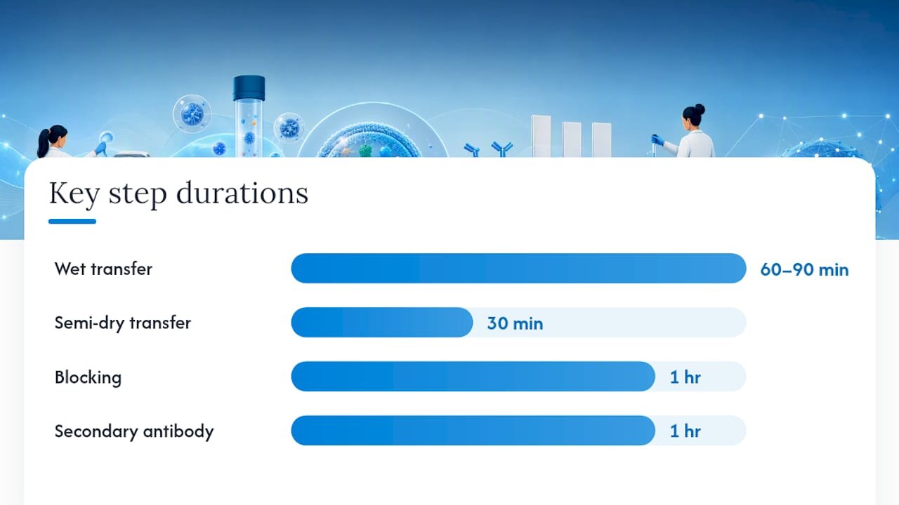
| Transfer | Wet or semi-dry, 100 V, 60–90 min (wet) | Semi-dry: 25 V, 30 min for most targets |
| Membrane | PVDF (activated in methanol) | Nitrocellulose acceptable for high-abundance targets |
| Transfer check | Ponceau S stain before blocking | Confirms even transfer; photograph and destain |
| Blocking | 5% milk or BSA in TBST, 1 hr RT | BSA preferred for phospho-targets |
| Primary antibody | Overnight at 4°C | See dilution guidance below |
| Secondary antibody | 1 hr RT | HRP or fluorescent conjugate |
Check transfer success by staining the membrane with Ponceau S immediately after transfer. A uniform stain across all lanes confirms even loading and transfer. Photograph the Ponceau image before destaining; it serves as your total-protein reference.
Detection and imaging
Chemiluminescence (ECL) is the most common detection method and works well for most EV markers. Infrared fluorescent detection (e.g., LI-COR Odyssey) offers a wider linear dynamic range and is preferable when you plan to quantify band intensities. For ECL, expose to film or a digital imager at multiple time points (30 sec, 2 min, 5 min) to capture both faint and strong bands without saturation.
Starting antibody dilutions for common markers:
- CD9, CD63, CD81: 1:500–1:1,000 (nonreducing conditions)
- TSG101: 1:1,000–1:2,000
- Alix: 1:500–1:1,000
- Calnexin: 1:1,000–1:2,000
- GM130: 1:500–1:1,000
QC embedded in the protocol: Before any quantitative comparison, run a two-fold dilution series of your EV lysate (e.g., 20 µg, 10 µg, 5 µg, 2.5 µg) on a single gel. Plot band intensity against protein input. Densitometry data commonly deviate from proportional linear models, and normalizing to a single loading-control protein can produce misleading results when linearity is not first confirmed. Only use data points that fall within the linear range for any quantitative claim.
Statistic callout: An analysis of critical factors for quantitative immunoblotting found that many factors, including sample preparation, detection method, and normalization strategy, influence linearity, and that diagnostic experiments such as dilution series are required before treating western-blot densitometry as quantitative.
What does each marker actually tell you on an exosome blot?
Tetraspanins: CD9, CD63, and CD81
These three are the most cited exosome western markers in the literature, and each has quirks. CD63 is the most specific for late-endosomal compartments but is also the most problematic to interpret: heavy N-glycosylation produces a broad smear from roughly 30 to 60 kDa rather than a discrete band. Do not mistake that smear for a failed blot. CD9 and CD81 tend to run closer to their predicted molecular weights (25–26 kDa) but still require nonreducing conditions for most commercial clones because their epitopes depend on disulfide-bond-maintained conformations.
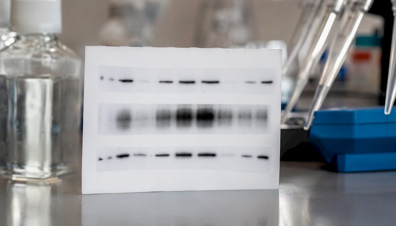
All three tetraspanins are enriched on EV surfaces, making them useful positive markers, but proteomics data show they appear across multiple EV subtypes, not exclusively in exosomes derived from MVB fusion. Treat their presence as evidence of EV enrichment, not proof of a specific biogenesis pathway.
TSG101 and Alix
TSG101 (~46 kDa) and Alix (~96 kDa) are cytosolic proteins recruited to MVBs during endosomal sorting. They are robust markers under standard reducing conditions, which makes them technically easier to detect than tetraspanins. TSG101 is particularly useful because it is relatively low-abundance in whole-cell lysate compared to EV fractions, so enrichment is visible. Alix runs at high molecular weight; if your transfer conditions are optimized for small proteins, you may under-transfer Alix. Extend wet transfer time to 90 minutes or use a gradient transfer protocol.
If your purification is poor, both TSG101 and Alix can appear in cell-debris fractions. Their presence in your prep is necessary but not sufficient evidence of EV purity.
Flotillin-1 and HSP70
Flotillin-1 (~48 kDa) marks lipid raft-associated membrane domains and is frequently detected in EV preps, but it is not specific to exosomes. HSP70 (~70 kDa) is a stress-inducible cytosolic chaperone that loads into EVs but is also elevated in stressed or rapidly dividing cell lines independent of EV content. Use both as supporting markers rather than primary evidence.
Negative controls: calnexin, GM130, and cytochrome c
- Calnexin (~90 kDa): ER-resident transmembrane protein. Its presence in your EV prep means ER membrane fragments co-isolated. This is a red flag for ultracentrifugation preps from cells with high ER content.
- GM130 (~130 kDa): Golgi matrix protein. Absence confirms your prep is free of Golgi contamination. Include for all cell-culture samples.
- Cytochrome c (~12 kDa): Mitochondrial marker. Relevant for cell-culture preps; rarely needed for plasma samples where mitochondria are not circulating.
For plasma and serum samples, lipoprotein particles co-isolate with EVs during ultracentrifugation and can inflate protein measurements. SEC fractionation reduces this, but always check your protein assay method: BCA assays can be biased by lipid content in biofluid EV preparations, so complement with Bradford or a chromatographic purity check.
Controls, normalization, and how to test linearity
Reliable densitometry starts with controls, not with the antibody.
Essential controls to include in every experiment:
- Blank lane: lysis buffer only, to check for background from the buffer itself
- Pre-isolation input sample: whole-cell lysate or unfractionated biofluid, to confirm marker enrichment in the EV fraction
- Positive control: a well-characterized cell lysate or purified exosome preparation with known marker expression
- Negative control lane: secondary antibody only (no primary), to assess non-specific background
- Cellular compartment markers: calnexin and GM130 as described above
Why single loading-control normalization often fails:
Dividing your target band intensity by a single housekeeping protein (e.g., beta-actin) assumes both proteins respond proportionally to changes in total protein loaded. That assumption frequently breaks down in western-blot data, particularly at the low protein amounts typical of EV samples. The housekeeping protein may saturate before your target does, or vice versa, producing a ratio that has no linear relationship to actual protein abundance.
Dilution-series protocol for linearity testing:
Prepare a two-fold serial dilution of your EV lysate across at least four points (e.g., 20, 10, 5, 2.5 µg). Run all four on a single gel alongside your experimental samples. After imaging, plot band intensity (y-axis) against protein input (x-axis) for both your target marker and your total-protein stain. A proportional linear relationship (R² > 0.95 is a reasonable acceptance threshold) confirms you are in the quantifiable range.
| Dilution point | Expected behavior | Action if nonlinear |
|---|---|---|
| Highest input (20 µg) | May saturate; exclude from linear range | Drop from quantification |
| Mid-range (10 µg) | Should fall in linear range | Use as anchor point |
| Low input (5 µg) | Confirm linearity holds | Include if R² > 0.95 |
| Lowest input (2.5 µg) | May fall below detection | Exclude if signal is noise-level |
Alternative normalization strategies:
Total-protein stains (Ponceau S, SYPRO Ruby, or stain-free gel technology) normalize to all proteins in the lane rather than one housekeeping protein, which is more robust for EV samples where housekeeping proteins may not be present at consistent levels. Spiked-in recombinant protein standards allow interpolation-based quantification: build a small standard curve on every membrane and read off absolute amounts rather than ratios. This approach avoids the pitfalls of ratio normalization when optical density data are nonlinear.
Pro Tip: A quick acceptance criterion for linearity: plot your dilution series, fit a linear regression, and check R². If R² falls below 0.95 or the highest-input point sits clearly above the regression line (saturation), discard that point and restate your quantitative claims using only the linear portion of the curve. Publish the dilution-series image in supplementary data so reviewers can judge your range.
Troubleshooting common exosome western blot problems
Weak or absent signal:
- Likely cause: insufficient protein input, over-diluted antibody, or failed transfer for membrane proteins.
- Fix: Ponceau-stain the membrane immediately after transfer to confirm protein is present. If Ponceau shows protein but antibody gives no signal, the antibody may require nonreducing conditions or a different blocking agent. If Ponceau shows no protein, the transfer failed.
- For biofluid samples with very low input, concentrate EV fractions further before lysis. A reversible Ponceau stain before antibody probing distinguishes lack of protein from transfer failure.
CD63 smear instead of a discrete band:
- This is expected behavior, not a problem. CD63 is heavily glycosylated and migrates as a broad smear between 30 and 60 kDa. If you need a cleaner band for quantification, treat the sample with PNGase F to remove N-glycans before running the gel.
High background:
- Likely cause: insufficient blocking, over-concentrated primary antibody, or cross-reactive secondary.
- Fix: increase blocking time to 2 hours, reduce primary antibody concentration, and add an extra wash step (3 × 10 min TBST). For plasma-derived samples, high background often reflects lipoprotein contamination; check whether SEC fractionation reduces it.
Inconsistent band intensity between replicates:
- Likely cause: variable protein loading, pipetting error at low volumes, or inconsistent lysis efficiency.
- Fix: always run a total-protein stain and normalize to it. For EV pellets, resuspend in a fixed volume and load a fixed volume rather than trying to equalize by BCA alone, especially for biofluid samples where BCA can be biased by lipid content.
Ladder bands absent from membrane after transfer:
- Likely cause: prestained ladder proteins did not transfer efficiently, or the membrane was not activated properly (PVDF requires methanol activation).
- Fix: confirm PVDF activation, check transfer buffer composition, and verify that the ladder lane was not at the gel edge where transfer is often uneven.
Quick QC checklist when a blot fails:
- Ponceau stain confirms protein transferred
- Antibody control lane (secondary only) rules out non-specific background
- Dilution series confirms you are in the linear range
- Check reducing vs. nonreducing conditions for tetraspanins
When a marker is genuinely absent after confirming transfer and antibody performance, that is a biological result worth reporting. Do not repeat the experiment indefinitely hoping for a different outcome; document the absence with appropriate controls and note it as a finding.
What orthogonal methods should you pair with western blot?
Western blot is a protein-presence assay, not a purity or biogenesis assay. Cross-platform workflows combining western blot with NTA, EM, single-EV immunodetection, and proteomics are the current standard for rigorous EV characterization.
Core orthogonal methods:
- Nanoparticle tracking analysis (NTA): Measures particle size distribution and concentration. Confirms that your prep contains particles in the 50–200 nm range expected for small EVs. Does not distinguish EVs from protein aggregates or lipoproteins, but size distribution combined with western data is far more informative than either alone.
- Transmission electron microscopy (TEM): Provides direct morphological evidence of vesicular structures. Cup-shaped morphology under TEM is a classic EV feature. Required by many journals for initial characterization.
- Single-EV immunodetection (e.g., ExoView): Captures individual particles on antibody-coated chips and detects surface markers at the single-particle level. Allows co-localization of CD9, CD63, and CD81 on the same vesicle, which western blot cannot do.
- LC-MS/MS proteomics: Provides an unbiased inventory of proteins in your prep. Published studies confirm EV marker presence by western blot and complement with LC-MS/MS for comprehensive validation.
- Flow cytometry (NATO-flow): Detects surface markers on individual particles above roughly 200 nm; less sensitive for small EVs but useful for larger microvesicle populations.
Recommended minimal method combinations:
- Cell-culture EVs: western blot + NTA + TEM. Add single-EV immunodetection if claiming subtype specificity.
- Plasma/serum EVs: SEC fractionation + western blot + NTA + single-particle immunodetection. The extra fractionation step reduces lipoprotein interference before any downstream assay.
MISEV/ISEV guidelines recommend reporting particle size, morphology, and protein markers as a minimum, and explicitly discourage claiming exosome-specific biogenesis without targeted mechanistic evidence. Build your characterization plan around those requirements from the start.
What actually matters when you validate exosome markers by western blot
Most labs treat the western blot as the endpoint. It should be the starting point for interpretation, not the finish line.
The markers themselves are well-established. CD9, CD63, CD81, TSG101, and Alix have been confirmed across hundreds of EV preparations, and the proteomics literature is clear that they co-enrich with small EVs. What the field underestimates is how much the quality of the blot determines whether those markers mean anything. A CD63 band in a prep that also shows strong calnexin signal tells you almost nothing useful about your EVs; it tells you the isolation failed.
The reproducibility problem in EV research is not primarily a marker-selection problem. It is a QC transparency problem. Dilution series and total-protein normalization data rarely appear in primary papers, even though they are the only way to know whether a band-intensity difference reflects biology or a pipetting error. Showing those data in supplementary figures is not extra work; it is the minimum standard for a quantitative claim.
When sample is limited, prioritize TSG101 and calnexin over the full tetraspanin panel. TSG101 is technically robust, enriched in EV fractions, and easy to detect under reducing conditions. Calnexin absence is the single most informative negative result you can report. If you have enough sample for only two antibodies, those are the two.
Mayflowerbio supports your exosome western blot workflow
Validated reagents and reliable sample characterization are where most EV western-blot workflows either succeed or stall. Mayflowerbio offers a direct path from sample to result: a catalog of validated antibodies covering the core exosome marker panel (CD9, CD63, CD81, TSG101, Alix, Calnexin, and more), each with documented western-blot performance data so you are not troubleshooting clone selection from scratch.

For sample characterization upstream of your blot, Mayflowerbio’s NTA size profiling service delivers particle size and concentration data that pair directly with your western results to meet MISEV reporting requirements. The exosome detection kits and isolation products cover the sample-prep stage, from ultracentrifugation-compatible concentration kits to SEC-based fractionation options for biofluid samples. To source validated antibodies or request NTA profiling for your next EV experiment, visit the Mayflowerbio exosomes product page and submit an inquiry directly through the site.
Sources
Key references used to build this guide:
- An analysis of critical factors for quantitative immunoblotting | Science Signaling
- Proteomic comparison defines novel markers to characterize heterogeneous populations of extracellular vesicle subtypes
- EV Characterization by Western Blot (protocol excerpt / methods)
FAQ
What are the standard positive markers for exosome western blots?
The standard positive panel includes tetraspanins CD9, CD63, and CD81 plus at least one ESCRT-associated cytosolic marker, either TSG101 or Alix, as recommended by ISEV/MISEV reporting guidelines.
Why does CD63 appear as a smear on western blot?
CD63 is heavily N-glycosylated, which causes it to migrate as a broad smear between roughly 30 and 60 kDa rather than a sharp band. PNGase F treatment before electrophoresis can sharpen the band if needed for quantification.
Do tetraspanin antibodies require nonreducing conditions?
Many CD63, CD81, and CD9 clones recognize disulfide-dependent epitopes and require nonreducing sample buffer (no DTT or beta-mercaptoethanol) to detect signal. Always check the antibody datasheet and test both conditions when using a new clone.
Is western blot alone sufficient to characterize exosomes?
No. Western blot confirms protein co-isolation but cannot prove vesicular morphology or biogenesis. MISEV guidelines recommend pairing western data with NTA for particle sizing and TEM for morphological confirmation at minimum.
How do you normalize western blot data for exosome samples?
Total-protein stains (Ponceau S or stain-free technology) are more reliable than single housekeeping proteins for EV samples, where housekeeping protein levels may be inconsistent. Always run a dilution series first to confirm you are working within the linear range of detection.




