

How to Calculate MOI for Viral Transduction Experiments
The multiplicity of infection formula is MOI = infectious particles added ÷ number of target cells. Flip it to solve for volume: Volume (mL) = (MOI × cell count) ÷ titer (infectious units/mL).
Say you have 500,000 cells and want an MOI of 3, using a lentiviral stock titered at 1 × 10⁸ TU/mL. Volume = (3 × 500,000) ÷ 1 × 10⁸ = 0.015 mL, or 15 microliters.
A few things determine whether that number actually works in your hands:
- Match units. PFU, TU, and vg are not interchangeable, and mixing them silently is one of the most common sources of failed reproducibility.
- Confirm your titer was generated on the same cell type you’re infecting, since permissiveness varies.
- Follow your institutional biosafety protocols and IBC approvals before handling any viral vector, regardless of how routine the calculation feels.
Key Takeaways
Accurate MOI calculation depends on a locally validated infectious titer, a cell count taken at the moment of infection, and clear separation between MOI_input and MOI_actual.
| Point | Details |
|---|---|
| Use the inversion formula | Volume (mL) = (MOI × cell count) ÷ titer solves for inoculum volume directly. |
| Poisson sets expectations | MOI 3 yields about 95% infected cells; MOI 5 yields about 99.3%. |
| Titer locally, not from the label | Vendor vg/mL can overstate infectious dose by a factor of 100 to 10,000 for AAV. |
| Adsorption time changes outcomes | MOI_actual can fall well below MOI_input if contact time or mixing is limited. |
| Reliable counting reduces variability | Mayflowerbio’s cell biology tools support accurate counts at the point of infection. |
This article is general information, not a substitute for advice from a qualified doctor. Consult a qualified healthcare professional about your own circumstances before acting on anything here.
Table of Contents
- MOI Calculation: Reading a Calculator’s Inputs Correctly
- What Percentage of Cells Get Infected at Each MOI?
- Dilution and Infection Protocol for a Target MOI
- How Do You Measure the Titer You’re Actually Calculating With?
- Common MOI Calculation Mistakes and How to Fix Them
- Sources
- FAQ
MOI Calculation: Reading a Calculator’s Inputs Correctly
Every MOI calculator, whether it’s a spreadsheet you built or a public tool, asks for the same five things: titer unit, total target cell count, desired MOI, infection volume constraint, and the minimum volume your pipette can reliably deliver. Miss any one of these and the output number is meaningless.
Here’s what each input actually does:
- Titer unit sets your entire calculation’s foundation. PFU/mL and TU/mL measure infectious activity directly; TCID50/mL estimates infectious dose from cytopathic effect; vg/mL counts genome copies, infectious or not.
- Cell count must reflect cells present at the moment of infection, not the number you originally plated.
- Target MOI depends on your goal: low for single-copy integration, high for near-universal infection.
- Volume constraint is dictated by your vessel (a 96-well plate holds far less than a T75 flask).
- Minimum pipettable volume matters because sub-microliter transfers introduce real error on most standard pipettes.
Working the algebra: if you need MOI 5 for 2 million cells and your stock is titered at 5 × 10⁸ PFU/mL, Volume = (5 × 2,000,000) ÷ 5 × 10⁸ = 0.02 mL, or 20 microliters, well above the practical pipetting floor.
One caveat worth flagging: if your only titer is TCID50 and you need PFU for the MOI calculation, multiply TCID50/mL by roughly 0.69 to approximate PFU/mL, a conversion that comes from the same Poisson math underlying the whole model.
Pro Tip: Build your calculator so titer unit is a required, unchangeable field, not a free-text box. Half of unit-mismatch errors happen because someone pasted a vg/mL number into a PFU/mL field without noticing.
What Percentage of Cells Get Infected at Each MOI?
The Poisson distribution answers this directly. The probability a given cell receives zero viral particles is P(0) = e^(−MOI), so the fraction of cells infected is 1 − e^(−MOI). This holds because viral particles distribute across a cell population randomly, not evenly, so some cells get hit multiple times while others get missed entirely, even at the same average MOI.
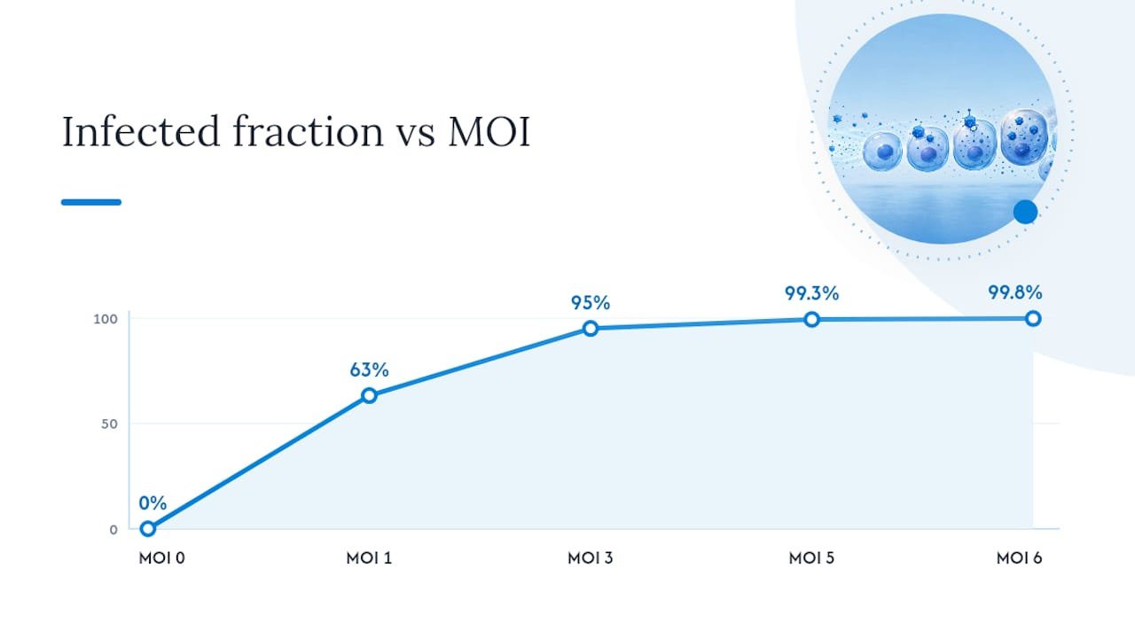
The rule of thumb researchers actually use: MOI 3 gets you to roughly 95% infected, and MOI 5 pushes past 99%. Diminishing returns kick in fast past that point.
There’s a catch, though. MOI_input, what you calculated and pipetted, is not MOI_actual, what actually adsorbed onto cells. Adsorption kinetics, contact time, and mixing efficiency all reduce actual particle uptake relative to the number added, and the gap can be substantial depending on your adsorption conditions. A rushed 15-minute incubation and a proper 1-hour rocking adsorption will not produce the same real infection rate, even with identical input MOI.
Dilution and Infection Protocol for a Target MOI
Getting from stock virus to a properly dosed well takes a specific sequence, and skipping steps is where most MOI errors creep in.
- Count cells at the moment of infection, not at plating. Cells proliferate between seeding and infection, and using a stale count skews your real MOI.
- Check viability with a standard exclusion dye; infecting compromised cells wastes virus and confounds your results.
- Thaw virus on ice and avoid refreeze/thaw cycles, which measurably degrade titer.
- Calculate dilution volume using the inversion formula. Example: your stock is 2 × 10⁹ TU/mL, you need MOI 1 for 1 million cells in a 500 microliter well volume. Required virus volume = (1 × 1,000,000) ÷ 2 × 10⁹ = 0.0005 mL, or 0.5 microliters, below most pipettes’ reliable range. Dilute the stock 1:100 in serum-free medium first, then pipette 50 microliters of the diluted stock instead.
- Add polybrene (commonly 4 to 8 micrograms per mL) for retroviral or lentiviral transductions to improve adsorption, but skip it for cell types sensitive to its cytotoxic effects.
- Incubate during adsorption, typically 4 to 24 hours depending on vector, then replace with fresh complete medium.
If your calculated volume still falls below the pipettable minimum after one dilution, split the diluted inoculum across replicate wells rather than forcing a single sub-microliter transfer.
Pro Tip: Serum can interfere with certain enveloped viruses’ entry efficiency. If your protocol calls for serum-free adsorption medium, don’t substitute your regular growth medium just because it’s on hand.
How Do You Measure the Titer You’re Actually Calculating With?
Vendor-stated titer is a starting point, not a number to build your MOI calculations on unvalidated. Local titering, done in your own cells under your own conditions, is what actually predicts your infection outcome.
- Plaque assays count PFU/mL by counting discrete plaques formed on a monolayer at limiting dilution. This works well for lytic viruses like adenovirus but takes days to read out.
- Fluorescence or flow-cytometry titering counts TU/mL by infecting cells with serial virus dilutions, then measuring the percentage expressing a reporter (GFP, for instance).
- TCID50 estimates infectious dose from the dilution causing cytopathic effect in 50% of replicate wells, useful when plaques don’t form cleanly.
- Vector genomes (vg/mL) count physical genome copies by PCR, regardless of whether those particles are infectious. AAV preparations commonly show vg to infectious-particle ratios ranging from 100:1 to 10,000:1, so vg-based MOI can dramatically overstate functional dose.
When calculating transduction titer by fluorescence, use wells showing under 40% positive cells. Above that threshold, multiple viral integrations per cell become common, and your TU/mL estimate skews artificially low.
Running a small dilution series on any new virus lot, rather than trusting a certificate of analysis, is the single habit that prevents the most downstream headaches.
Common MOI Calculation Mistakes and How to Fix Them
Most failed or inconsistent transductions trace back to one of a handful of repeatable errors.
- Using vendor vg/mL as if it were infectious titer. Fix: titer locally with a fluorescence or plaque assay before calculating MOI for a critical experiment.
- Counting cells at plating instead of at infection. Fix: recount immediately before adding virus, especially for fast-dividing lines.
- Assuming MOI_input equals MOI_actual. Fix: extend or standardize adsorption time, and treat MOI as a starting estimate, not a guarantee, particularly near threshold doses.
- Ignoring pipetting minimums. Fix: pre-dilute concentrated stocks in serum-free medium before the final addition step.
- Skipping unit labels in your notebook. Fix: write PFU, TU, or vg next to every titer number, every time.
Pro Tip: Keep a running log of titer method, lot number, and adsorption conditions for every virus stock. Six months later, that log is the only thing that explains why one experiment’s MOI 3 behaved differently from another’s.
A Note on Reproducibility
The gap between a clean MOI calculation and a reproducible result usually comes down to what you didn’t write down. Cell passage number, exact medium composition, adsorption time, and which titering method generated your denominator all shift outcomes in ways a formula alone won’t catch. Log them every time, even when the experiment “worked,” because the failed repeat six weeks later is when you’ll wish you had.
— Alina
Reagents and Cell Counts That Keep MOI Calculations Honest
Bad inputs produce bad MOI numbers no matter how careful the math is, and the two most common culprits are inconsistent cell counts and virus stocks that were never properly re-titered in-house. Mayflowerbio’s cell biology tools support accurate, at-time-of-infection cell counting, which removes one entire category of MOI drift before it starts.

For labs running frequent transductions, Mayflowerbio’s viral transduction and bioprocessing line covers reagents built for consistent, lot-to-lot adsorption performance, and the cell biology checklist walks through QC steps worth building into any titering workflow. If your last MOI series gave you results you couldn’t explain, start by checking whether your cell counts and reagent lots were the variable. Browse Mayflowerbio’s cell biology product line to see what fits your current protocol.
Sources
- Fluorescence titering assay — Addgene
- MOI (Multiplicity of infection) Calculator — Gera Tools
- Poisson
FAQ
What Does MOI of 0.1 Mean?
An MOI of 0.1 means you’re adding one infectious particle for every ten cells on average, which the Poisson model predicts will infect roughly 9.5% of the cell population. Researchers use this low range for single-copy integration work where minimizing multiple hits per cell matters more than infecting every cell.
What MOI Should I Use for Near-Complete Infection?
MOI 3 typically infects about 95% of cells, and MOI 5 pushes that to roughly 99.3%, based on the standard Poisson calculation. Going much higher than MOI 5 usually wastes virus for diminishing returns.
Can I Use Vector Genome (vg) Counts to Calculate MOI?
You can, but vg-based MOI often overstates the functional dose because it counts all genome copies, infectious or not, and AAV preparations can carry vg to infectious-particle ratios as high as 10,000:1. Use a PFU or TU-based titer whenever the experiment’s outcome depends on precise dosing.
Why Is My Actual Infection Rate Lower Than My Calculated MOI Predicted?
MOI_actual, the particles that truly adsorbed onto cells, is often lower than MOI_input because adsorption kinetics and limited contact time reduce real uptake. Extending adsorption time or improving mixing typically closes some of that gap.
How Do I Titer a New Virus Stock Before Calculating MOI?
Mayflowerbio’s cell biology checklist outlines the QC steps that keep this process consistent across lots.




