
Stop Blood PCR False Negatives for Labs with BSA or Tolerant Polymerases
Hemoglobin and immunoglobulin G (IgG) are the two dominant PCR inhibitors in blood, and anticoagulants like heparin and EDTA make things worse when handling goes wrong. If your Cq values are drifting or your positive partitions are dropping, the fastest wins are switching to an inhibitor-tolerant polymerase or running a proper extraction and cleanup step. Adding bovine serum albumin (BSA) to the reaction buys you a quick, low-effort mitigation while you sort out the bigger fix.
TL;DR:
- Hemoglobin and its breakdown products cause enzyme inhibition and fluorescence quenching, often leading to delayed or failed amplification in blood samples.
- Immunoglobulin G binds to DNA and blocks primer annealing, especially in plasma samples, raising Cq values without necessarily stopping amplification.
- The anticoagulant used during blood collection, particularly heparin and EDTA, significantly impacts PCR success due to direct inhibition or magnesium chelation, respectively.
- Fluorescence quenching from heme compounds can produce weak or flat signals, making positive reactions indistinguishable from negatives unless probed with less susceptible chemistries.
- Employing inhibitor-tolerant enzyme formulations, adding BSA, optimizing collection protocols, and choosing appropriate extraction methods are key to mitigating blood-derived PCR inhibitors effectively.
Table of Contents
- What Causes PCR Inhibition in Blood Samples?
- Which Blood Components Are the Biggest PCR Inhibitors?
- How Do Blood Inhibitors Change qPCR and dPCR Results?
- Best Practices for Blood Collection and Anticoagulant Selection
- Extraction and Cleanup Methods That Remove PCR Inhibitors
- Choosing Inhibitor-Tolerant Polymerases and Reaction Facilitators
- Troubleshooting Checklist for PCR Amplification from Blood Samples
- About This Guide and Mayflowerbio’s Role in Inhibitor Mitigation
- Where Purity Trades Off Against Sensitivity
- Mayflowerbio’s Reagents for Inhibitor-Prone Blood Workflows
- Sources
- FAQ
What Causes PCR Inhibition in Blood Samples?
Blood is not a clean template. It carries proteins, metabolic byproducts, and iron-containing molecules that interfere with amplification at nearly every stage of the PCR reaction, and each one does it differently. Understanding which mechanism is at play in your assay determines which fix actually works, because a facilitator that neutralizes hemoglobin will not touch an IgG problem.
Polymerase inhibition by hemoglobin and heme. Hemoglobin, and its breakdown products heme and hematin, bind directly to DNA polymerase and interfere with nucleotide incorporation. Iron released from heme groups can also generate reactive oxygen species that damage the enzyme’s active site over the course of thermal cycling. This is why samples with even mild hemolysis often show delayed or absent amplification, especially at low template concentrations where the enzyme has little margin for error.
Template binding by IgG. IgG works through a completely different route. Purified plasma IgG binds single-stranded genomic DNA, and researchers found it can block primer annealing or physically obstruct polymerase access during the early cycles when template is still double-stranded and vulnerable. One study found that mixing plasma with ssDNA-agarose beads reduced this inhibitory activity, which tells you the IgG is binding nucleic acid directly rather than just gumming up the enzyme. Oddly, heating purified IgG together with target DNA increased inhibition in the same experiments, a detail that matters if your protocol includes a hot-start denaturation step before the enzyme is active.
Fluorescence quenching by heme and hematin. This is a detection problem, not an amplification problem, and it is the one most likely to trick you into thinking your assay failed when it actually worked. Heme and hematin absorb and quench fluorescent signal, which suppresses passive reference dyes like ROX and dsDNA-binding dyes like EvaGreen or SYBR Green. Published experiments have shown quenching that scales with hemoglobin concentration, meaning you can have a positive reaction sitting right in your tube with a flat, uninterpretable curve on the instrument.
Chelation and low-molecular-weight inhibitors. EDTA chelates magnesium, and magnesium is the cofactor your polymerase needs to catalyze the reaction, so residual EDTA carried over from a collection tube can quietly kill amplification even at trace concentrations. Bilirubin, bile salts, and iron salts like FeCl3 round out the list of small-molecule troublemakers, and they tend to show up more in hemolyzed or icteric samples.
The mechanisms break down into four broad categories worth keeping on a lab wall chart:
- Enzyme inhibition: hemoglobin, heme, and iron byproducts interfere with polymerase function directly.
- Template interference: IgG binds single-stranded DNA and blocks primer or polymerase access.
- Signal masking: heme and hematin quench fluorescent dyes without necessarily stopping amplification.
- Cofactor depletion: EDTA and related chelators strip magnesium away from the reaction.
A review synthesizing inhibition mechanisms across qPCR, digital PCR, and massively parallel sequencing makes the point that no single mitigation strategy addresses all four categories at once. That is the single biggest mistake labs make when troubleshooting: they assume more BSA or a longer extraction will fix everything, when the actual problem might be signal masking that no amount of extra facilitator will solve.
Which Blood Components Are the Biggest PCR Inhibitors?
Not every inhibitor in blood carries equal weight. Some are potent enough to shut down amplification at micromolar concentrations, while others only matter when handling goes badly wrong. Here is the practical inventory, ranked roughly by how often they cause real trouble in the lab.
Hemoglobin, heme, and hematin top the list because they come from the most abundant cellular component in blood, red blood cells, and they attack PCR on two fronts at once: direct polymerase inhibition and fluorescence quenching. Even samples that look only mildly hemolyzed can carry enough free hemoglobin to shift your Cq values. This is the inhibitor most likely to be behind an unexplained assay failure when nothing else in your protocol has changed.
Immunoglobulin G is the major plasma-derived inhibitor. Because IgG is present in every plasma sample regardless of hemolysis status, it is arguably the more persistent threat for assays that rely on plasma rather than whole blood, and researchers have identified IgG as a major inhibitor of diagnostic PCR that raises Cq values in a concentration-dependent manner, with measurable Cq shifts reported around 190 μM.
Lactoferrin, purified from leukocytes, is a lesser-known but real contributor. It is an iron-binding protein that interferes with polymerization, and it shows up specifically in samples with a higher white blood cell fraction, such as buffy coat preparations or leukocyte-rich extracts.
Anticoagulants deserve their own line item because they are the one inhibitor category a lab actually controls at the point of collection:
- Heparin is a potent, direct polymerase inhibitor and one of the hardest anticoagulants to work around once it is in the sample.
- EDTA chelates magnesium and drops polymerase activity unless the reaction buffer’s Mg2+ is adjusted to compensate.
- Citrate is generally the gentler option of the three for downstream molecular work, though it is not universally inert either.
Other contributors round out the picture: bilirubin and bile salts from icteric or lipemic samples, residual leukocyte genomic DNA that competes for reagents in low-template reactions, and, outside clinical settings, humic-like environmental contaminants that can carry over from field or forensic collection.
How Do Blood Inhibitors Change qPCR and dPCR Results?
Inhibition rarely announces itself. It usually just looks like a failed or weak result, and the trick is telling the difference between “there wasn’t enough template” and “there was plenty of template but something blocked it.”
In quantitative PCR, the clearest signature is a rightward shift in Cq, sometimes accompanied by a change in amplification efficiency that flattens the exponential phase of the curve. In digital PCR, inhibition shows up as a drop in the percentage of positive partitions relative to what you would expect from your input concentration, since dPCR is less sensitive to efficiency loss but still vulnerable to complete reaction failure in individual partitions.
Fluorescence quenching deserves special attention because it can mimic a true negative. Heme and hematin absorb light in wavelengths that overlap with common reporter and reference dyes, so a reaction can amplify target DNA successfully while producing a curve too dim to call positive. Passive reference dyes like ROX are particularly susceptible, and experiments have documented quenching that tracks directly with hemoglobin and hematin concentration in the sample, alongside reduced EvaGreen and SYBR-based signal under the same conditions.
Some assay formats hold up better than others under inhibitor load:
- Probe-based assays (TaqMan-style) tend to be less vulnerable to dye quenching than dsDNA-binding dye assays, because the probe’s reporter dye is only released upon hydrolysis, which changes the signal dynamics compared to a dye that binds any double-stranded product.
- Dye-based assays (SYBR, EvaGreen) are more exposed to quenching artifacts since the entire signal depends on dye intercalation into amplified product.
- Multiplex assays compound the problem, because inhibition rarely affects every target equally. A weaker amplicon in a multiplex panel can drop below detection while a stronger one still calls positive, producing a misleading partial result.
- Thermolabile or engineered enzyme systems built for inhibitor tolerance generally outperform standard hot-start polymerases when the matrix is difficult.
Pro Tip: Build an internal positive control (IPC) spike into every batch of blood-derived samples. If your IPC shows delayed Cq or dropout alongside your target, you are looking at inhibition, not a true negative, and no amount of re-running the same extraction will fix that.
Running external standards alongside spike-in IPCs is the most reliable way to catch inhibition before it corrupts a result set, and it costs almost nothing compared to re-collecting a sample.
Best Practices for Blood Collection and Anticoagulant Selection
The single most controllable inhibitor variable in your entire workflow is the anticoagulant choice made at the moment of blood draw. Get this step wrong and no downstream cleanup fully compensates.
- Choose EDTA or citrate over heparin whenever the protocol allows it. Heparin is a direct, potent PCR inhibitor and one of the more difficult anticoagulants to neutralize once present, while EDTA’s inhibitory mechanism, magnesium chelation, can be compensated for by adjusting the reaction buffer’s Mg2+ concentration.
- Match your matrix to your assay. Plasma gives you a cleaner cellular background but a higher relative IgG load; whole blood carries the full hemoglobin burden plus leukocyte-derived lactoferrin; serum sits in between and avoids anticoagulant inhibition entirely but introduces clotting-related variability. Pick based on which inhibitor category your assay tolerates better.
- Minimize the delay between draw and processing. Extended storage at room temperature promotes hemolysis, which directly increases free hemoglobin and heme in the sample. Refrigerate promptly and process within the window your protocol validates for.
- Handle the tube gently. Rough mixing, forceful aspiration, and repeated freeze thaw cycles all promote hemolysis. This sounds trivial, but it is one of the most common preventable sources of inhibitor load in routine sample handling.
- For field and forensic collection, weigh sensitivity against inhibitor risk. Dried blood spots and buccal swabs reduce liquid-handling hemolysis risk but concentrate inhibitors like hemoglobin and heme into a smaller elution volume, which can paradoxically raise local inhibitor concentration even as total sample volume drops.
None of these steps require new equipment. They require discipline at the collection bench, which is exactly why they get skipped under time pressure and why inhibition problems so often trace back to the earliest step in the workflow rather than the PCR reaction itself.
Extraction and Cleanup Methods That Remove PCR Inhibitors
Once inhibitors are in the sample, extraction and cleanup are where you actually remove them rather than just working around them. The right method depends on how much inhibitor load you are fighting and how much target you can afford to lose in the process.
Silica-column and magnetic-bead extraction remain the workhorses for blood matrices. Both chemistries bind nucleic acid selectively while inhibitory proteins and small molecules wash through, and magnetic-bead protocols in particular scale well for automation in high-throughput clinical or forensic labs. The tradeoff is that more aggressive wash steps that improve inhibitor removal can also reduce yield on already low-copy targets.
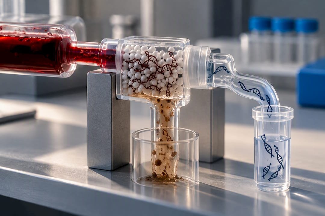
Adsorption and affinity-based approaches target specific inhibitors rather than nucleic acid generally. Experiments have shown that mixing plasma with ssDNA-agarose beads reduces IgG’s inhibitory activity by binding the protein directly, which is a useful secondary cleanup step when IgG is the suspected culprit rather than hemoglobin.
Enzymatic and chemical pretreatments offer another angle. Proteinase K digestion ahead of extraction helps break down inhibitory proteins including IgG and residual hemoglobin-binding complexes, and specific heme-removal chemistries exist for samples with heavy hemolysis where quenching is the dominant concern rather than enzyme inhibition.
- Serial dilution is the fastest rapid mitigation available. Diluting the template reduces inhibitor concentration proportionally, but it also dilutes your target, so it only works when starting copy number is high enough to tolerate the loss.
- Spin-column cleanup post-extraction offers a middle ground: faster than full re-extraction, more effective at inhibitor removal than dilution alone.
- Combination approaches (proteinase K digestion followed by silica-column extraction) tend to outperform any single method when inhibitor load is severe.
Pro Tip: Never assume a protocol change that fixes inhibition also preserves sensitivity. Re-run your limit of detection (LOD) and precision checks after any extraction or cleanup modification. A method that clears hemoglobin beautifully but quietly doubles your LOD has just traded one problem for another.
Purification stringency and target sensitivity pull against each other in almost every blood-derived workflow. The goal is not maximum inhibitor removal. It is the minimum cleanup that gets your assay back to validated performance.
Choosing Inhibitor-Tolerant Polymerases and Reaction Facilitators
Enzyme and buffer choice can eliminate the need for aggressive extraction entirely, and this is often the fastest fix available when a sample is already extracted and you are troubleshooting on the bench.
- Look for engineered or fusion polymerases built for difficult matrices. Comparative studies show real differences in blood tolerance between polymerase formulations, and some engineered enzymes or fusions with ssDNA-binding domains allow amplification directly from a much higher percentage of whole blood than standard hot-start enzymes tolerate.
- Add BSA as a first-line facilitator. Experiments testing 0.4% BSA in reaction buffer showed increased amplification tolerance across tested enzyme systems, and it is cheap, fast to trial, and compatible with most existing protocols without reformulation.
- Consider ssDNA-binding proteins like gp32 where IgG-mediated template binding is the suspected mechanism. Facilitators that occupy or shield single-stranded regions can reduce the competition IgG creates for primer annealing sites.
- Run a formal inhibitor-tolerance panel before committing to a production protocol. Spike serial concentrations of hemoglobin, purified IgG, and heparin into a control template, test your candidate polymerase and facilitator combinations across that gradient, and plot the resulting Cq shift or percent-positive change in dPCR. This tells you exactly where your protocol breaks down instead of leaving you to guess after a batch fails in production.
Pro Tip: Do not run your tolerance panel with only one inhibitor at a time. Real blood samples carry hemoglobin, IgG, and residual anticoagulant simultaneously, and their effects are not always simply additive. A polymerase that handles high hemoglobin fine might still fail against a moderate hemoglobin plus IgG combination.
Dye selection matters here too. If you are already fighting quenching from hemolysis, pairing a facilitator-enhanced polymerase with a probe-based chemistry avoids stacking two vulnerabilities, dye masking and enzyme inhibition, on top of each other in the same reaction.
Troubleshooting Checklist for PCR Amplification from Blood Samples
When a blood-derived PCR run fails or looks suspicious, work through this sequence rather than randomly changing variables:
- Check your controls first. Every batch should include a positive template control, an inhibition control (an IPC spiked into the actual sample matrix), and an extraction blank. If the IPC shows delayed Cq or dropout, you have confirmed inhibition rather than a true negative.
- Try rapid, low-effort mitigations before re-extracting. Dilute the template two- to five-fold, add BSA to the reaction, or swap in an alternate polymerase known for blood tolerance. Any one of these can resolve mild to moderate inhibition in minutes.
- If the problem persists, re-extract using a different chemistry. Move from a silica column to magnetic beads, or add an adsorption-based cleanup step targeting IgG specifically if plasma is your matrix.
- Validate before you trust the fix. Re-check LOD, precision, and robustness on the revised protocol. A fix that resolves inhibition but quietly shifts your detection limit is not actually a fix, and the trade-off between purification stringency and sensitivity loss is worth re-checking every time you change a step.
Keep this sequence in a shared lab document. The labs that lose the least time to inhibition problems are the ones that run it as a habit, not as a last resort after three failed batches.
About This Guide and Mayflowerbio’s Role in Inhibitor Mitigation
This guide draws on peer-reviewed inhibition mechanism studies to give lab teams a working reference, not a substitute for validating your own assay. Suppliers provide molecular biology reagents for handling difficult matrices: PCR mixes, polymerases designed for challenging samples, extraction kits, and cleanup beads suited to blood-derived samples. If your lab is running an inhibitor-tolerance panel or troubleshooting a persistent blood-matrix problem, reaching out for technical support can save a batch of samples rather than losing them to a guess. Support teams can field exactly these kinds of protocol questions from research and clinical labs.
Where Purity Trades Off Against Sensitivity
Every lab eventually faces the same tension: clean your sample harder and you risk losing the low-copy target you actually care about; clean it less and inhibition creeps back in. There is no universal answer, and pretending otherwise sets teams up to over-purify routine samples that never needed it.
My honest take is that most labs default to aggressive cleanup out of habit rather than necessity, when a facilitator-enhanced polymerase and a well-designed IPC would have caught the problem earlier and cheaper. Routine inhibition controls should be a standard line item in every assay validation, not an afterthought added after a batch fails. Reserve full extraction and cleanup for samples where direct PCR with a tolerant enzyme has already been tried and demonstrably falls short.
— Alina
Mayflowerbio’s Reagents for Inhibitor-Prone Blood Workflows
If hemoglobin, IgG, or anticoagulant carryover keep showing up in your Cq data, the fix usually lives in your reagent choice before it lives in a new extraction protocol. Mayflowerbio stocks other PCR reagents built for exactly this kind of difficult matrix work, alongside qPCR mixes suited to both dye and probe chemistries.

For labs weighing dye quenching risk against probe-based reliability, the HOT FIREPol Probe Universal qPCR Mix sidesteps the hemoglobin quenching problem that affects dsDNA-binding dyes, while the HOT FIREPol EvaGreen qPCR Mix Plus remains a solid option for capillary platforms where inhibitor load is already well controlled. Mayflowerbio also carries antibodies for downstream validation work when IgG interference is part of your assay design. Current pricing for these reagent lines is available directly on the product pages. If you are setting up an inhibitor-tolerance panel and need a small validation sample or protocol input from the technical support team, reach out through the Mayflowerbio products catalog to get started.
FAQ
What Are Examples of PCR Inhibitors Found in Blood?
The main ones are hemoglobin, heme, and hematin from red blood cells, plus immunoglobulin G from plasma. Lactoferrin from leukocytes, anticoagulants like heparin and EDTA, and small molecules such as bilirubin and bile salts also contribute.
What Does PCR Inhibition Mean for Bloodwork Results?
It means a component in the blood sample is blocking or masking amplification, producing a false negative or an artificially delayed Cq rather than an accurate reflection of the actual target concentration. This is why inhibition controls and IPC spikes matter for any clinical or forensic result derived from blood.
How Do You Remove PCR Inhibitors From a Blood Sample?
Silica-column or magnetic-bead extraction removes most inhibitors during standard nucleic acid purification, and adsorption approaches using ssDNA-agarose beads specifically target IgG. Faster in-reaction options include diluting the template, adding BSA, or switching to an inhibitor-tolerant polymerase.
Is EDTA a PCR Inhibitor?
Yes, but through a specific and correctable mechanism: EDTA chelates magnesium, and magnesium is the cofactor your polymerase needs to function. Adjusting the reaction buffer’s Mg2+ concentration typically compensates for residual EDTA carried over from collection tubes.




