

How to Run a Pseudovirus Neutralization Assay: Protocol and Validation
A pseudovirus neutralization assay measures antibody-mediated blockade of viral entry using single-cycle pseudotyped particles, reporting potency as an ID50 or NT50 that converts to IU/mL against the WHO International Standard 20/136. Researchers favor it because:
- It runs at BSL-2, unlike live-virus plaque reduction neutralization tests (PRNT) that need BSL-3 containment.
- Readouts come from luciferase or GFP reporters, giving quantitative, high-throughput data in a single infection cycle.
- Titers are comparable across labs once normalized to a shared international standard.
Key Takeaways
A pseudovirus neutralization assay delivers PRNT-comparable neutralization data at BSL-2 by measuring single-cycle entry inhibition and reporting it as an ID50/NT50 traceable to WHO IS 20/136.
| Point | Details |
|---|---|
| Assay principle | PBNAs measure single-cycle entry inhibition, not multi-cycle replication, so results can diverge from PRNT at high antibody concentrations. |
| Backbone choice | VSV yields faster, higher titers but risks residual helper virus; lentiviral/MLV systems give more stable reporter expression. |
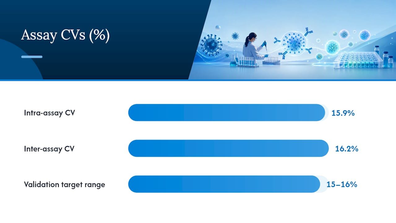
| Validation benchmarks | Target intra-/inter-assay CVs near the 15 to 16% range and define LOD using negative serum panels (mean plus 1.96 SD). |
| Standardize inputs | Fix pseudovirus dose and convert NT50 to IU/mL using WHO IS 20/136 for cross-lab comparability. |
| Reagent sourcing | Mayflowerbio supplies GFP-reporter VLPs, neutralizing monoclonal controls, and cell counting consumables to support consistent PBNA setup. |
This article is general information, not a substitute for advice from a qualified doctor. Consult a qualified healthcare professional about your own circumstances before acting on anything here.
Table of Contents
- What Does a Pseudovirus Neutralization Assay Actually Measure?
- What Materials Do You Need to Produce and Titrate Pseudovirus?
- How Do You Set Up the Neutralization Plate and Readout?
- How Do You Analyze and Validate the Data?
- Luciferase or Fluorescent Reporter: Which Fits Your Assay?
- Why Do Neutralization Results Vary Between Runs?
- What Reagents Does Mayflowerbio Offer for PBNA Workflows?
- Get Reagents and Support for Your Neutralization Assay Setup
- Sources
- FAQ
What Does a Pseudovirus Neutralization Assay Actually Measure?
A pseudovirus-based neutralization assay (PBNA) captures one thing: how well an antibody or serum sample blocks a pseudotyped particle from entering a target cell during a single round of infection. That single-cycle design is the whole point. It isolates entry inhibition from the downstream replication, spread, and cytopathic effects that complicate live-virus assays like PRNT.
Two backbone systems dominate the field. Vesicular stomatitis virus (VSV) pseudotyping, typically using a glycoprotein-deleted VSV-ΔG core, packages fast and yields high titers within 24 to 48 hours. Its main liability is residual helper virus carryover, which can produce false-positive infection signals if not controlled during production. Lentiviral (HIV-based) or MLV pseudotyping systems take longer to produce but integrate reporter genes more stably and tend to show lower background, making them a common choice for labs running extended screening campaigns.
- VSV: fast turnaround, high titer, watch for residual helper virus.
- Lentiviral/MLV: slower production, stable reporter expression, lower background noise.
- Both correlate well with live-virus neutralization in most spike-mediated entry contexts, per comparative work on standardized PBNA correlation with PRNT.
- Divergence from PRNT tends to show up at high antibody concentrations or when the antibody acts through a non-entry mechanism, since a single-cycle assay cannot capture post-entry effector functions.
Pro Tip: If you’re screening a monoclonal panel and one candidate shows a flat, non-sigmoidal curve, don’t assume assay failure first. Check whether that antibody’s mechanism is entry-independent — Fc-mediated effector functions won’t show up in a PBNA at all.
What Materials Do You Need to Produce and Titrate Pseudovirus?
Building a reliable pseudovirus stock starts with the right plasmid set and a disciplined harvest schedule. For lentiviral systems, you need a packaging plasmid (Gag-Pol), an envelope/spike-expression plasmid, and a transfer plasmid carrying your reporter gene, transfected into HEK293T cells. VSV systems substitute a VSV-ΔG backbone plasmid rescued in a producer line already expressing the desired glycoprotein.
- Transfect producer cells (HEK293T for lentiviral; BHK-21 or HEK293T for VSV) with a validated ratio of packaging, envelope, and reporter plasmids.
- Harvest supernatant at 48 to 72 hours post-transfection for lentiviral systems, or 24 to 30 hours for VSV, since VSV cytopathic effects set in fast.
- Clarify by low-speed centrifugation or 0.45 µm filtration, then concentrate by ultracentrifugation or PEG precipitation if higher titers are needed.
- Titrate the clarified stock on your target cell line before freezing working aliquots.
Reporter choice matters as much as backbone. Firefly luciferase remains the workhorse for high-throughput screening because of its wide dynamic range, while GFP suits imaging and flow-based readouts where you need single-cell resolution.
- Plasmids: packaging, envelope/spike, reporter transfer vector.
- Transfection reagent: PEI or a lipid-based reagent, titrated per producer cell batch.
- Titration method: RLU-based endpoint dilution, converted to TCID50-equivalent units.
- Target MOI: 0.1 to 0.5 for most neutralization formats, or an empirically determined TCID50 that yields signal 20 to 50 fold over background.
How Do You Set Up the Neutralization Plate and Readout?
Plate layout decides whether your data holds up under scrutiny. Edge wells run warmer and evaporate faster in standard incubators, so keep virus-only and cell-only controls away from the outer rows whenever plate real estate allows.
- Prepare serial dilutions of serum or antibody, typically 3-fold across 6 to 9 points, starting around a 1:20 or 1:40 initial dilution for serum panels.
- Pre-incubate diluted samples with a fixed pseudovirus dose for 30 to 60 minutes at 37°C to allow antibody-virus binding before cells are added.
- Add target cells at a seeding density suited to your line: HEK293T-hACE2 cells run well around 1 to 2 x 10^4 cells per well in a 96-well format; Huh7 and Vero derivatives may need slightly higher densities for consistent monolayer formation.
- Include a virus control (no antibody), a cell control (no virus), a negative human serum panel, and a positive reference standard such as a validated monoclonal or WHO IS 20/136 dilution series.
- Incubate 48 to 72 hours for luciferase readouts (lentiviral systems) or 18 to 24 hours for VSV-based GFP systems, since VSV cycles faster.
- Read luminescence on a plate luminometer with integration time and gain settings matched to your reporter’s dynamic range, or acquire flow cytometry / imaging data with consistent gating thresholds across the run.
Pro Tip: Co-expressing TMPRSS2 alongside ACE2 in your target cell line changes entry kinetics for spike-pseudotyped particles. If your neutralization curves look unexpectedly shallow, confirm which entry pathway your target cells actually favor before blaming the serum panel.
How Do You Analyze and Validate the Data?
Fit your dose-response data with a four-parameter logistic regression to derive ID50 or NT50, the sample dilution or antibody concentration producing 50% neutralization relative to virus-only control. GraphPad Prism handles this natively through its nonlinear regression module, and most labs fix the top and bottom plateaus when signal saturates rather than letting the fit float freely.
Converting raw NT50 values to IU/mL against WHO IS 20/136 is what makes your data comparable to another lab’s, or to a multi-site trial dataset. Without that conversion step, an NT50 of 320 in your assay means nothing outside your own plate.
- Determine LOD/LLOQ from a negative serum panel using the mean plus 1.96 standard deviations, run separately per serum matrix (human versus animal sera behave differently).
- Track intra- and inter-assay %CV across repeated runs; a validated SARS-CoV-2 PBNA reported intra-assay CV near 15.9% and inter-assay CV near 16.2%, a useful benchmark range for your own acceptance criteria.
- Report R² values alongside raw signal ranges so other labs can judge curve-fit quality, not just the final NT50 number.
Assays with intra-/inter-assay CVs in the 15 to 16% range and clearly defined LOD cutoffs from negative panels are considered well-controlled by current validation literature, giving you a concrete target when setting your own QC thresholds.
Luciferase or Fluorescent Reporter: Which Fits Your Assay?
Reporter choice shapes throughput, sensitivity, and what kind of biological question you can actually answer. Luciferase delivers the widest dynamic range and slots cleanly into 96 or 384-well HTS pipelines, but substrate handling introduces its own variability if injector timing or reagent age isn’t controlled. GFP and RFP reporters cost you some throughput but let you resolve single-cell entry events by imaging or flow cytometry, which matters when you need to confirm that a neutralization signal reflects blocked entry rather than a downstream artifact.
| Reporter | Best for | Main tradeoff |
|---|---|---|
| Luciferase | High-throughput plate-based screening | Substrate handling affects linearity |
| GFP | Imaging, flow cytometry, single-cell kinetics | Lower throughput per run |
| RFP | Multiplexed imaging alongside GFP markers | Dimmer signal in some cell lines |
Running an orthogonal readout, luciferase for screening and flow cytometry for confirmation. This approach catches cases where a compound or antibody affects reporter expression itself rather than viral entry.
Why Do Neutralization Results Vary Between Runs?
Residual VSV helper virus is the most common source of false positives in VSV-pseudotyped systems. Optimizing producer MOI, tightening harvest timing, and adding a clarification or ultracentrifugation step reduces contaminating helper virus, and some labs pre-treat stocks with anti-VSV neutralizing antibody before assay use as an extra safeguard.
- If EC50 or R² values drift between runs, retitrate your target cells. Seeding density shifts of even 20% can move the entire dose-response curve.
- Animal sera (mouse, ferret) show higher nonspecific background than human sera, so set LOD cutoffs per species rather than applying one blanket threshold.
- Fix your pseudovirus input dose across runs, in TCID50 or IU/mL, and include a shared reference serum in every plate to anchor inter-run comparisons.
Pro Tip: Keep a running log of RLU values for your virus-only control across every plate you run. A slow downward drift over several months usually means your working pseudovirus aliquot is losing titer in the freezer, not that your antibodies are getting weaker.
What Reagents Does Mayflowerbio Offer for PBNA Workflows?
Running a pseudovirus neutralization assay well depends on reagent consistency as much as protocol discipline. Mayflowerbio supplies SARS-CoV-2 virus-like particles with GFP reporters for wild-type spike work, along with variant-adapted XBB.1.5 GFP-reporter particles for labs tracking circulating strains.
- Pseudotyped particles and VLPs across multiple spike variants for imaging or flow-based readouts.
- Neutralizing monoclonal antibodies suited as positive reference controls in your dilution series.
- Cell counting slides and consumables that support accurate MOI calculation and consistent seeding density across runs.
- US-based technical support for protocol questions and reagent selection.
Pro Tip: A miscounted cell stock is one of the quietest sources of assay drift. Pairing a fixed pseudovirus input with a reliable cell counting method removes one full variable from your troubleshooting list before you even start the neutralization curve.
A Lab-Tested Approach to Running Consistent PBNAs
Batch your pseudovirus preps in bulk and freeze single-use aliquots rather than producing fresh stock for every run. Run-to-run variability drops noticeably once you stop reintroducing production variance into every experiment. Track titer, RLU range, and %CV on a rolling basis, and re-prepare stock once CV creeps outside your established range rather than waiting for a failed run to tell you. When results feed into a clinical or regulatory claim, pair the PBNA with an orthogonal assay, PRNT where feasible, rather than leaning on a single readout.
Get Reagents and Support for Your Neutralization Assay Setup
Mayflowerbio gives researchers a direct path to the pseudotyped particles, reporter constructs, and antibody controls a PBNA actually needs, without piecing together reagents from five different vendors with inconsistent lot documentation.

That matters most when you’re trying to hit the CV benchmarks and LOD standards this article just walked through. Inconsistent reagent sourcing is one of the quietest ways an otherwise well-designed assay drifts out of validation range between runs. Mayflowerbio’s catalog covers neutralizing monoclonal antibodies for your positive controls, cell biology consumables for seeding accuracy, and infectious disease reagents for building out your panel, all backed by US-based technical support if you need help matching a reagent to your specific pseudovirus system. If you’re setting up or re-validating a PBNA workflow, start by browsing the antibody catalog for a positive reference standard that fits your target virus.
Sources
- Quantitative and standardized approaches for PBNA normalization (PMC8641955)
FAQ
What Does a Positive SARS-CoV-2 Antibody Test Mean?
A positive antibody test indicates prior exposure to the virus or vaccination, but it does not by itself confirm neutralizing capacity. A pseudovirus neutralization assay is needed to determine whether those antibodies actually block viral entry.
What Is a Pseudovirus?
A pseudovirus is a non-replicating viral particle, typically built on a VSV or lentiviral core, that displays a target virus’s surface glycoprotein without carrying its full genome. It allows entry-blocking antibodies to be studied safely at BSL-2 instead of BSL-3.
What Are the Different Types of Neutralization Assays?
The two main formats are pseudovirus-based neutralization assays (PBNAs), which use single-cycle pseudotyped particles at BSL-2, and plaque reduction neutralization tests (PRNT), which use replicating live virus at BSL-3. PBNAs are faster and more scalable; PRNT remains the traditional reference method for many pathogens.
What Is a Virus Neutralization Test?
A virus neutralization test measures the ability of antibodies in a serum or plasma sample to block viral infection of target cells, typically reported as a titer such as ID50 or NT50. Both live-virus PRNT and pseudovirus-based assays fall under this category, differing mainly in biosafety level and turnaround time.
Are ID50, IC50, and NT50 the Same Measurement?
They describe closely related but not identical endpoints. ID50 and NT50 typically refer to the sample dilution giving 50% neutralization, while IC50 usually refers to an absolute antibody concentration, so matching the metric to how your data will be compared downstream matters when reporting results.




