How to Prevent Mycoplasma Contamination in Cell Culture
You prevent mycoplasma contamination by controlling what enters your cell culture room (new cell lines, serum, media and people), by handling cultures with strict aseptic technique, and by testing on a fixed schedule so a breach is caught in days, not months. No single step is enough. Mycoplasma does not cloud the medium, and the usual antibiotics do not touch it, so prevention is a system you run, not a thing you notice.
This guide is written for lab managers, core facility staff and bench scientists who want a plan they can put into a standard operating procedure this week.
TL;DR
- Quarantine every incoming cell line in a dedicated incubator until it has tested negative, and test again after it has grown in antibiotic-free medium.
- Test all lines by PCR or DNA staining at least monthly, and treat the result as a documented quality control record, not an ad hoc check.
- Keep penicillin and streptomycin out of routine culture. They do not act on mycoplasma and can hide a problem.
- Buy serum, media and cell lines from suppliers who certify them mycoplasma-free, and filter or test what you cannot certify.
- When a culture turns positive, discard it unless it is irreplaceable, then decontaminate the hood and incubator before anything else goes in.
Table of Contents
- Why mycoplasma is a different kind of contaminant
- Where mycoplasma comes from
- Step 1: Control what comes into the lab
- Step 2: Make aseptic technique a habit, not a reminder
- Step 3: Stop relying on antibiotics
- Step 4: Test on a schedule
- Our perspective: treat it as quality control, not cleanup
- What to do when a culture tests positive
- Answers to common worries
- Building the supply side of your QC
- A one-page SOP outline you can adapt
- FAQ
- Sources
Why mycoplasma is a different kind of contaminant
Bacteria and fungi announce themselves. The medium turns cloudy, the pH indicator shifts, and you throw the flask away before lunch. Mycoplasma does none of this. Reviews of the topic describe cultures that look normal while the organisms grow, and labs often notice only when growth rate, morphology or assay results drift, by which point the contamination may have been present for weeks or months.
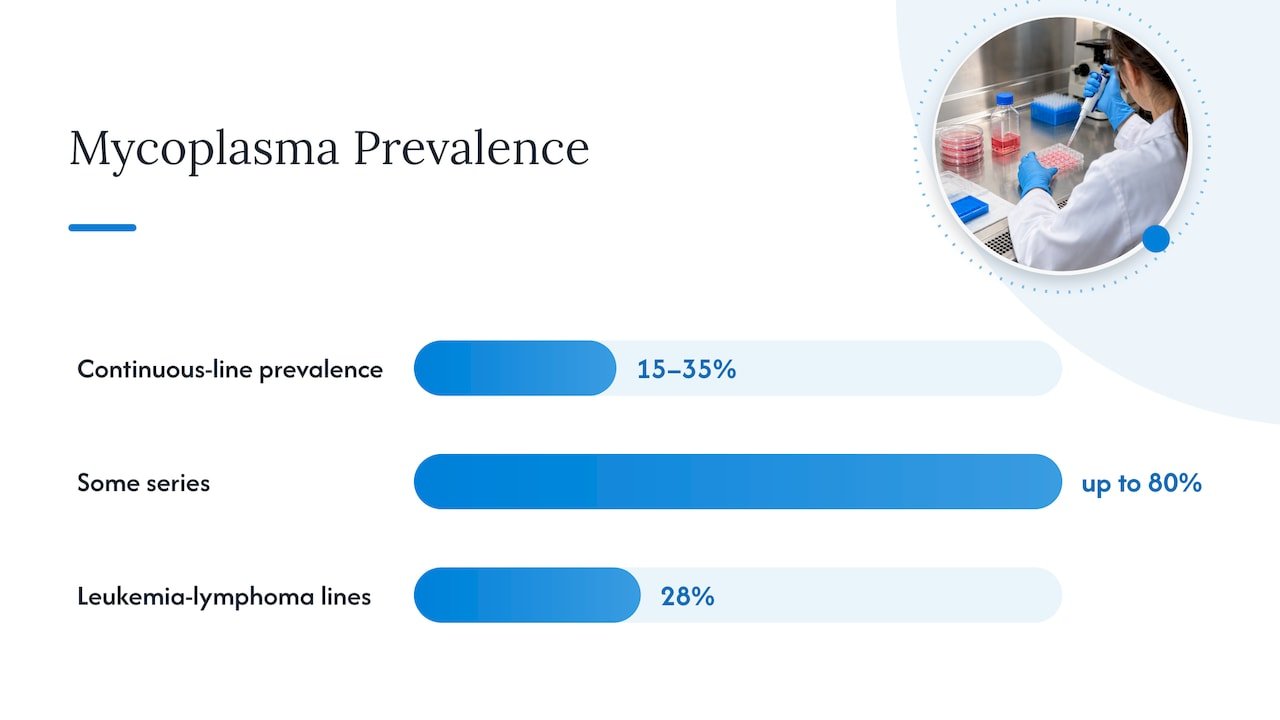
The scale of the problem explains why it deserves its own protocol. A classic review by Hans Drexler and Cord Uphoff puts prevalence in continuous cell lines at roughly 15 to 35 percent, with some series reporting up to 80 percent. The same work reports titers of 10^6 to 10^8 organisms per mL in infected cultures, with 100 to 1,000 organisms attached to each host cell. A study of more than 460 human leukemia-lymphoma lines found 28 percent of cultures positive.
The consequences are not cosmetic. The same 2023 review notes that infection triggers global changes in gene expression and chromatin state in host cells. For anyone running drug discovery, transfection, epigenetics or RNA-seq work, a contaminated line can quietly invalidate months of data. That is the real cost to budget against.
Where mycoplasma comes from
Prevention gets easier once you know the entry routes. The literature points to the same short list.
- Other cell lines. A review of prevention and detection states that a single contaminated culture is enough to endanger every other culture in the lab, and it recommends testing every line obtained from an outside source.
- Operators. Aerosols and particles generated during handling spread the organisms from flask to flask. Pipetting habits and shared reagent bottles do the rest.
- Animal-derived reagents. The FDA names animal serum products and the culture facility environment as the two major sources, particularly with open culture systems.
- Equipment and surfaces. Incubator water pans, hood surfaces, centrifuge buckets and water baths all carry organisms between runs if they are not on a cleaning schedule.
Drexler’s own conclusion in the same review is that prevention is far more cost-effective than eradication, and that efforts are in vain if cells go back into a contaminated environment or are handled with poor practice. In other words, good cell culture practice is the primary defense.
Step 1: Control what comes into the lab
Quarantine new cell lines
Treat every incoming vial as positive until proven otherwise, whether it came from a commercial repository, a collaborator down the hall or a lab across the country. The Salk Institute’s cell technologies core states that all new, approved lines are cultured under quarantine until they test clean for mycoplasma. That is a sound model for any lab.
A workable quarantine setup looks like this:
- A dedicated incubator, or at minimum a dedicated shelf with its own tray and labeling, that never holds established stocks.
- A set order of work: established lines first, quarantined lines last, with the hood wiped down between.
- Separate media aliquots for quarantined cultures, so a shared bottle cannot carry anything across.
- Written release criteria: which test, how many negatives, and who signs off.
One test on arrival is not enough. The NCATS quality control team describes testing after cells have been passaged for a period in antibiotic-free medium, so any suppressed organisms can climb to detectable levels. Low-level contamination can slip past PCR if cell density or sample handling is poor, so plan on several independent negatives before release.
Source reagents with care
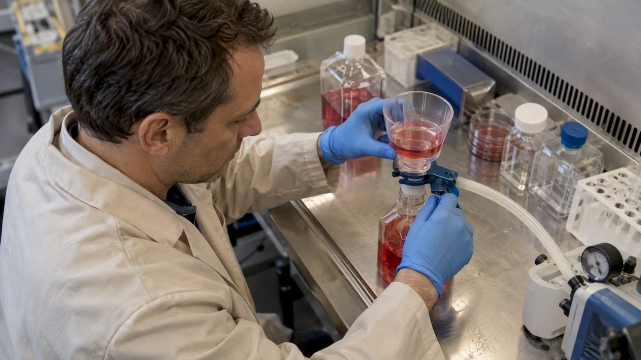
Serum is the classic carrier. Use serum and other animal-derived components from suppliers who certify them mycoplasma-free, and keep the certificates with your lot records. Reviews of prevention also advise testing medium components, especially serum, for sterility before use. According to the prevention literature, most culture media cannot be autoclaved, so filtration matters, and a low pressure differential of 5 to 10 psi is less likely to force mycoplasma through a membrane than 20 psi or more. If you filter in house, check your pump settings.
For cell lines, the same logic applies. Buy from repositories and vendors who authenticate and test their stocks, and ask for the testing method and date. If a line comes from another lab without a result, it goes into quarantine like any other.
Step 2: Make aseptic technique a habit, not a reminder
Most of the guidance on technique is unglamorous, and that is the point. The safety manual guidance from Florida State University lists the basic steps for reducing contamination: use good aseptic technique, reduce accidents, keep the laboratory clean, monitor routinely, use a frozen repository strategically and use antibiotics sparingly.
Turn those into daily behavior:
- Work with one cell line at a time in the hood, and clear the surface between lines.
- Never share media bottles between lines or between people. Aliquot into single-use volumes.
- Use filtered tips and sterile, individually wrapped serological pipettes. Do not pour from flask to flask.
- Keep the hood uncluttered. Airflow works only when the front grille and back slots are clear.
- Disinfect with 70% ethanol before and after each session. Guidance on protecting cells also covers wiping equipment with ethanol or decontaminating with UV where appropriate.
- Clean incubators on a calendar. Drain and sanitize water pans, wipe shelves and replace trays at a fixed interval, and log it.
- Minimize open handling. Closed systems and sealed flasks reduce exposure, which matters because the FDA flags open culture systems as a risk.
- Do not talk over open vessels, and tie back hair and wear a clean lab coat dedicated to the culture room.
These measures also keep bacterial and fungal contamination down, so the payoff is not limited to mycoplasma.
Step 3: Stop relying on antibiotics
It is tempting to add penicillin and streptomycin to every bottle as insurance. For mycoplasma, the insurance does not pay out. The Drexler review explains that mycoplasmas are resistant to most antibiotics commonly used in cell culture, and the Florida State guidance recommends using antibiotics sparingly. Routine use suppresses the bacteria that would otherwise warn you that technique has slipped, which gives a false sense of security.
A practical policy:
- Run routine culture antibiotic-free whenever the line and the experiment allow it.
- If a protocol needs antibiotics, use them for a defined purpose and period, not by default.
- Always include an antibiotic-free passage period before a mycoplasma test, as described in the quarantine step above.
If your lab has been on antibiotics for years, expect a few surprises when you stop. A line that suddenly shows bacterial growth was telling you something about your technique.
Step 4: Test on a schedule
Prevention without testing is a guess. Testing is the only way to know your controls work.
How often should you test?
The research summary behind this article points to testing all cell lines by PCR or DNA staining at least monthly, as outlined in a 2023 review of prevention, diagnosis and eradication. Add tests at these moments:
| Moment | Why |
|---|---|
| On arrival and after antibiotic-free passaging | Catches contaminated incoming lines |
| Before freezing a new master or working bank | Protects your repository |
| Monthly, for every line in active culture | Limits how long an infection can spread unnoticed |
| Before key experiments, publications or grant submissions | Protects data integrity |
| Before sharing a line with another lab | Protects collaborators |
| After any suspected breach or unexplained drift in growth or assay results | Confirms or rules out the cause |
The NCATS team recommends treating mycoplasma testing as a core part of quality control, with a negative result required for data reproducibility, grant submission and line sharing. Put it in the SOP, assign an owner and keep the records.
Choosing a method
Three approaches dominate.
- Culture-based assays. Sensitive, but they can take up to 28 days, which the FDA notes is impractical for products with limited dating periods.
- DNA staining. Fluorescent staining of host cell cultures is quick and inexpensive, but it needs an experienced eye and indicator cells.
- PCR and qPCR. Fast, sensitive and easy to batch. For regulated products, the FDA gene therapy CMC guidance recognizes PCR-based assays as a rapid, sensitive and specific option, provided the sponsor validates sensitivity and specificity.
If you work on cell therapy or gene therapy products, note that the FDA expects mycoplasma testing on cell banks, intermediate stages and final product. Check the FDA somatic cell therapy CMC guidance for what applies to your submission.
A few testing tips that save repeat work:
- Sample supernatant from cultures at high density that have been in the same medium for at least a few days, without a medium change right before.
- Include a positive and a negative control in every run, and an inhibition control so a failed reaction is not mistaken for a negative.
- Record lot numbers for primers, master mix and controls alongside the result.
- Retest any ambiguous result rather than interpreting it.
If you build PCR assays in house, you can find general guidance in our molecular assay setup guide for optimized PCR workflows and an overview of types of molecular biology assays.
Our perspective: treat it as quality control, not cleanup
We supply reagents and tools to labs working in cell biology, molecular biology and drug discovery, and the pattern we see in the literature and in customer conversations is consistent: the labs that suffer most are the ones that treat mycoplasma as an occasional emergency. The labs that stay clean treat it as a routine measurement, like checking a balance or calibrating a pipette.
Three things we would put at the top of any lab’s plan:
- Make one person accountable for the schedule. Monthly testing slips when it belongs to everyone.
- Keep a cell line register. Source, date received, passage number, last test date and result, storage location. When a positive appears, the register tells you in minutes which lines shared an incubator or a bottle.
- Link testing to downstream work. If a line has not tested negative recently, it should not go into an expensive assay. Mycoplasma changes the biology you are trying to measure, and cell authenticity is the companion problem. Our cell biology checklist and guide to reliable cell culture techniques cover the QC habits that go with it.
We would rather see a lab spend an afternoon writing a clear SOP than spend a quarter repeating experiments. Prevention is cheap by comparison.
— Alina
What to do when a culture tests positive
Even good labs get hits. How you respond decides whether it stays one flask or becomes a lab-wide problem.
- Isolate immediately. Stop work on the affected line and move it out of shared incubator space.
- Check neighbors. Test every line that was handled in the same session, hood or incubator, plus any line that received shared media.
- Discard if you can replace it. Prevention reviews recommend discarding positive cultures, and the research summary above advises autoclaving them to prevent spread. Go back to a frozen, tested stock if you have one.
- Treat only what you cannot replace. Treatment of valuable, unique cultures is feasible, as the same review notes, but follow it with repeated testing, including after an antibiotic-free period, to confirm elimination.
- Decontaminate. Clean the hood, incubator, water bath and any shared equipment thoroughly, and replace opened media and reagents that touched the culture.
- Find the source. Review the register, serum lots and recent arrivals. If you cannot identify the route, tighten the control that seems weakest.
For irreplaceable lines, talk to your institution’s biosafety officer or cell culture core before choosing a treatment route, because they will know the approved protocols for your facility.
Answers to common worries
What does mycoplasma look like in culture?
Usually nothing. The medium stays clear and the pH indicator does not change. Clues are subtle: slower growth, odd morphology, reduced transfection efficiency or inconsistent assay results. That is why testing, not observation, is your defense.
Can a lab ever be mycoplasma-free for good?
Not as a permanent state, because new lines, reagents and people keep arriving. You can keep the rate of incidents very low and the time to detection short, which is what the quarantine and monthly testing do.
Building the supply side of your QC
Testing needs reliable reagents, and prevention needs consistent counting, separation and authentication. If you are standardizing your workflow, our catalog covers cell counting and cell separation products, qPCR and RT-qPCR reagents and other PCR reagents for labs that run their own PCR-based checks. For authentication alongside mycoplasma control, we also offer a karyotype analysis service. Prices are quoted on request, so contact us through the site with your line list and workflow, and browse the full product range if you want to see everything in one place.
A one-page SOP outline you can adapt
If you want to leave this page with something concrete, draft your SOP under these headings:
- Scope and responsibility. Which lines and rooms are covered, and who owns testing and the register.
- Receiving and quarantine. Dedicated incubator, order of work, antibiotic-free passaging, number of negatives needed for release.
- Routine handling. One line at a time, no shared bottles, ethanol before and after, cleaning calendar for hoods and incubators.
- Reagent control. Approved suppliers, certificates on file, serum lot testing or filtration parameters.
- Testing. Method, schedule, controls, acceptance criteria and where results are recorded.
- Positive result response. Isolation, neighbor testing, discard or treatment decision, decontamination and sign-off.
- Review. Quarterly look at the register: how many incidents, how long until detection, which control failed.
The quarterly review is the step most labs skip, and it is the one that turns a document into a working defense.
FAQ
How often should I test for mycoplasma?
At least monthly for every line in active culture, using PCR or DNA staining, according to a 2023 review of prevention and eradication. Also test on arrival, before banking, before key experiments or publication, and any time growth or assay results change unexpectedly.
Do penicillin and streptomycin prevent mycoplasma?
No. Mycoplasmas are resistant to most antibiotics commonly used in cell culture. Routine use can mask contamination by suppressing other bacteria, so use antibiotics sparingly and test after an antibiotic-free passage period.
Will mycoplasma ever go away on its own?
No. Once established it keeps growing and can spread to other cultures through aerosols, shared reagents and handling. Discard the culture where you can replace it, or treat it under a validated protocol and retest repeatedly.
What are the first signs of mycoplasma?
There is often no visible sign. Watch for slower growth, changes in morphology, falling transfection efficiency or assay results that no longer reproduce. Because these signs are non-specific, rely on scheduled testing.
Sources
- Prevention, Diagnosis and Eradication of Mycoplasma Contamination in Cell Culture (PMC, 2023)
- Mycoplasma contamination of cell cultures: Incidence, sources, effects, detection, elimination, prevention (Drexler and Uphoff, 2002)
- Mycoplasma contamination of cell cultures, full text (PMC)
- Prevention and Detection of Mycoplasma Contamination in Cell Culture (PMC)
- Keeping it clean: cell culture quality control at NCATS (PMC)
- Prevention of mycoplasma contamination in leukemia-lymphoma cell lines (PubMed)
- Understanding and Managing Cell Culture Contamination (Florida State University)
- Salk Institute Cell Technologies and Engineering Core resources
- FDA guidance: CMC information for human gene therapy INDs
- FDA guidance: CMC information for human somatic cell therapy INDs
Recommended
- Cell Culture Master Reliable Techniques for Consistent Lab Result
- Prevent Cell Clumping in Single Cell Prep: DNase Rescue in 15–30 Min
- Fix Lentiviral Transduction in Primary and Suspension Cells: 0.1–10 MOI
- T-Store® Tissue Storage and Transportation Medium with Antibiotic (Amphotericin B and nanomycopulitin)