Exosome-Depleted FBS: Methods, Trade-Offs and Controls
Exosome-depleted FBS is fetal bovine serum that has been processed to remove bovine extracellular vesicles (EVs), so the vesicles you isolate from your cultured cells are not mixed with vesicles from the serum. You need it whenever you collect EVs, exosomes or their RNA and protein cargo from conditioned medium. It is never perfectly clean, it can slow your cells down, and you should verify it in your own hands. This guide covers how depletion works, which method removes the most, what it costs your cultures, and which controls keep your data defensible.
TL;DR
- Standard FBS is full of bovine EVs and RNA. If you collect vesicles from conditioned medium, that background can pass for your own signal.
- Ultracentrifugation (about 100,000 to 120,000g for 18 hours) is the common method, but it never reaches zero. In one head-to-head study, ultrafiltration removed more particles and protein.
- Depleted serum often slows proliferation and shifts differentiation, because growth factors and hormones leave with the vesicles. Test your cell line before committing.
- Always run a non-conditioned medium control, and confirm depletion by nanoparticle tracking rather than trusting an “EV-free” label.
Table of Contents
- Why standard FBS contaminates EV work
- How exosome-depleted FBS is made
- What depletion does to your cells
- Verify depletion yourself
- Terminology: exosomes, EVs and what you can claim
- Do you need it at all?
- Serum, plasma and other sources
- Our perspective
- Where Mayflower Bioscience fits
- Research use only: a note on therapy claims
- A practical checklist before your next collection
- FAQ
- Sources
Why standard FBS contaminates EV work
Fetal bovine serum is the default growth supplement for mammalian cell culture, and it carries its own population of vesicles. Those vesicles transfer functional molecules, including RNA, between cells. A study on depletion protocols showed that serum EVs contain functional, RNA-containing material and that depletion protocols matter for eliminating it. If you then isolate vesicles from the medium your cells conditioned, you recover two populations: the ones your cells made and the ones that came in the bottle.
The practical consequences are easy to picture:
- A vesicle RNA-seq or small RNA qPCR experiment can pick up bovine sequences or serum-derived signal that you attribute to your cells.
- A particle count by nanoparticle tracking will include serum particles, inflating your apparent yield.
- A functional assay, such as treating recipient cells with your isolated EVs, can be confounded by active serum vesicles.
The review of serum influence on in vitro EV analyses describes the three classic options that have been used since the 2006 proposal by Thery and colleagues: serum-free media, 1% bovine serum albumin in place of whole FBS, or FBS that has gone through an EV-depletion protocol. Exosome-depleted FBS is the third route, and it is the one most labs reach for because it keeps a familiar serum-based culture system.
How exosome-depleted FBS is made
There are two broad families of approaches, and the choice affects both purity and what is left in the serum afterward.
Ultracentrifugation
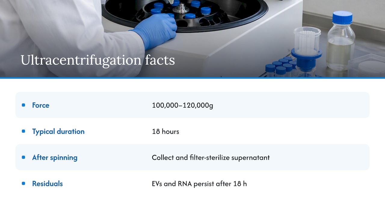
The industry-standard method is ultracentrifugation at 100,000 to 120,000g, typically for 18 hours, after which the supernatant is collected and filter-sterilized. It is simple in concept and every EV lab has the equipment. The downsides are time, the need for a high-speed rotor, careful handling of the pellet and supernatant interface, and the fact that serum is viscous, which reduces sedimentation efficiency compared with a dilute buffer.
Even the gold-standard protocol leaves something behind. The study of depletion protocols found that residual EVs and associated RNA persist after 18 hours at 120,000g, which can act as a confounding variable in sensitive experiments. So “depleted” means reduced, not removed.
Ultrafiltration
Ultrafiltration passes serum through a membrane with a defined molecular weight cutoff, so vesicles and large complexes are retained or removed by size, depending on how the workflow is set up. A comparison of depletion protocols using umbilical cord mesenchymal stromal cells reported that ultrafiltration achieved 89.77% particle depletion and 99.11% protein depletion, against 78.27% and 70.41% for ultracentrifugation (p < 0.01 and p < 0.0001 respectively).
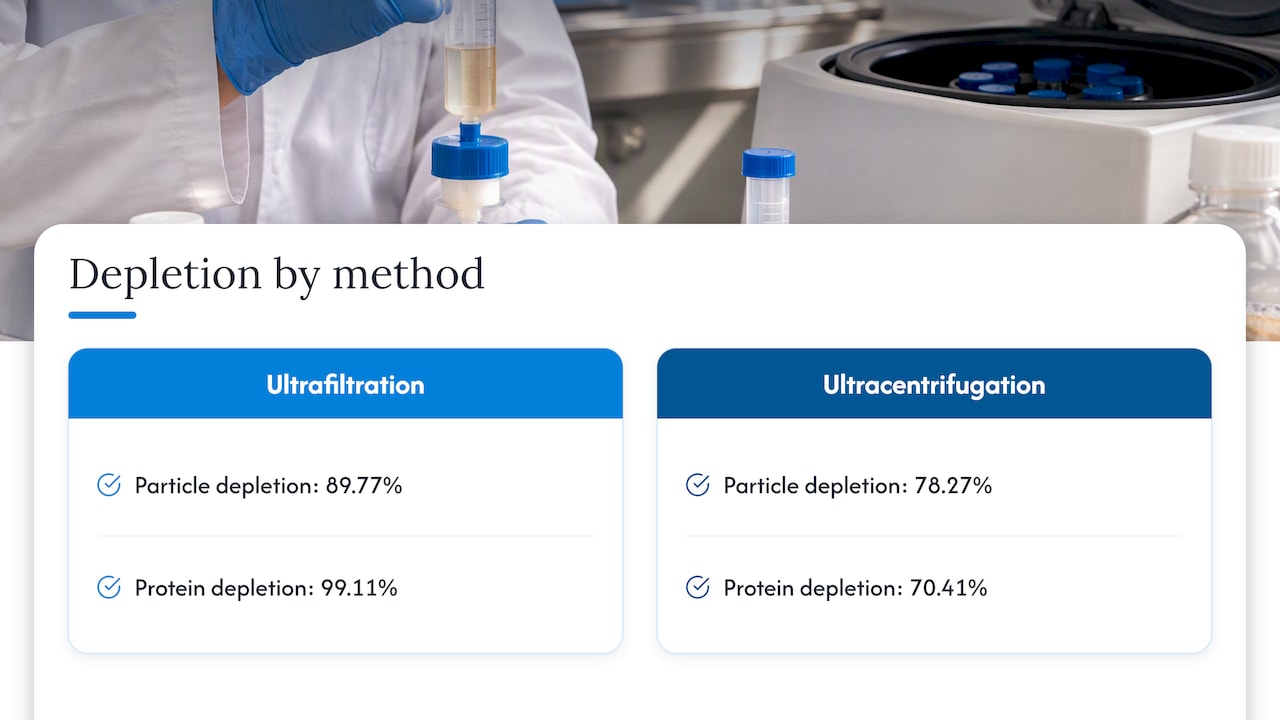
Read those numbers carefully. Particle depletion is what you want for an EV experiment. Protein depletion of 99% sounds even better until you remember that serum protein is what feeds your cells. A method that strips almost all protein is likely to leave a medium that cells struggle in, which brings us to the cost side.
Commercial and kit-based options
Most labs also have the option of buying ready-made depleted serum or using a depletion kit on regular FBS. Ready-made serum saves a day or more of centrifugation and removes operator variability, but lots differ, and the claims on the label are the supplier’s, not yours. Kits shorten the protocol, but you still need to show they work for your serum lot and your downstream readout. Whichever you choose, treat the product as a reagent to be qualified, not a guarantee.
Method comparison
| Approach | What it removes | Main advantage | Main caution |
|---|---|---|---|
| Ultracentrifugation, 100,000 to 120,000g, ~18 h | Most larger vesicles; less efficient in viscous serum | Standard, equipment already in most EV labs | Residual EVs and RNA remain; long run time |
| Ultrafiltration | Higher particle and protein removal in one reported comparison | Greater particle depletion in that study | Heavy protein loss may affect growth |
| Purchased depleted serum | Varies by supplier and lot | Saves time, less handling | You still have to verify depletion |
| Serum-free or 1% BSA medium | Avoids serum vesicles entirely | No bovine EV background | Many cell lines do not tolerate it well |
What depletion does to your cells
Depletion is not a neutral cleanup. The chondrogenic study in an osteoarthritis mouse model and related work describe a double-edged effect: exosome-depleted FBS reduces background from bovine vesicles, but it frequently lowers proliferation and alters differentiation potential compared with native FBS, because growth factors and hormones can be removed along with the vesicles.
This matters most for:
- Primary cells and stem or stromal cells, where phenotype is sensitive to the culture environment. The umbilical cord MSC study cited above looked at exactly this: how the depletion protocol itself influenced phenotype, immunomodulation and particle release.
- Differentiation protocols, where a lost factor can change the outcome without any obvious sign in the dish.
- Slow-growing lines, where a modest drop in proliferation turns into a very small harvest of conditioned medium.
Practical ways to keep cells healthy
A PLOS One study on EV isolation combined cell medium modifications with ultrafiltration and size-exclusion chromatography. Its approach points to a common practical fix: moving cells to a lower percentage of depleted serum, around 1%, in an advanced culture medium that supplies albumin, transferrin and insulin. Many researchers find this keeps proliferation better than standard medium with 10% depleted serum.
A workable adaptation plan looks like this:
- Grow cells in normal serum to a healthy, sub-confluent state.
- Wash twice with PBS so serum carryover is minimal.
- Switch to your depleted-serum medium for a test run in parallel with a control flask in standard medium.
- Compare viability, doubling time and the morphology you care about over at least one full collection window.
- Only then scale up conditioned medium collection.
If cells fail the test, step down the serum percentage, or move to a defined, serum-reduced formulation, and re-test. Document the change, because reviewers will ask.
Verify depletion yourself
The most useful habit in this area is skepticism toward the label. The MISEV2023 guidelines stress that serum-derived EVs can persist and co-isolate with cell-derived EVs, and that non-conditioned medium controls are essential to tell serum contaminants apart from your actual target. Researchers are advised to confirm depletion efficiency in their own setting instead of assuming that a commercial “EV-free” product is devoid of particles.
A minimal verification set:
- Unconditioned medium control. Put the complete medium, with depleted serum, in an empty flask in the incubator for the same time as your cells. Process it through the same isolation workflow. Whatever you recover is background.
- Particle profiling. Run the depleted serum, the unconditioned medium and your isolate by nanoparticle tracking to compare size distribution and particle concentration. Our NTA size profiling service exists for labs that do not have an instrument in house.
- Marker checks. Use western blotting or a comparable approach for positive and negative markers, and keep in mind that bovine and human proteins can cross-react with antibodies. Our exosome western markers reference is a practical place to start.
- Lot tracking. Record serum lot numbers and keep one tested lot in reserve for a whole project.
A note on concentration units
Particle concentration from NTA is normally reported as particles per milliliter, and many labs add particles per cell or per microgram of protein to normalize across conditions. Whatever unit you choose, state the sample volume, dilution and instrument settings, because the same preparation can give different numbers on different setups. The point is to compare like with like: depleted serum background against your sample, in the same units.
Terminology: exosomes, EVs and what you can claim
The search term is “exosome-depleted FBS”, and suppliers use it constantly, but the field has moved on. The MISEV2023 guidance notes that “exosome” is a biogenesis-specific term that is often misused. Researchers are encouraged to say extracellular vesicles unless a specific subcellular origin can be shown, since most isolation methods yield a mix of particles. That is why you also see “EV-depleted FBS” and “FBS EV depletion” in papers.
In your methods section, describe what you actually did and measured: the serum, the depletion method with its g-force and duration, and the characterization. A reviewer cares more about that than about the word on the bottle.
Do you need it at all?
Not every experiment calls for depleted serum. A fair rule of thumb:
| Situation | Depleted FBS likely worth it? |
|---|---|
| Isolating EVs from conditioned medium for RNA, proteomics or functional assays | Yes, or an alternative that removes serum EVs |
| Downstream readouts where a small background is acceptable and controlled | Maybe, with unconditioned medium controls |
| Routine culture, passaging, cell line maintenance | No, use standard serum |
| Fragile or primary cells that fail without full serum | Consider a short serum-free collection window instead |
A common compromise is to grow cells in regular serum, wash thoroughly, and then collect conditioned medium for 24 to 48 hours in serum-free or depleted medium. This limits the time cells spend under stress while keeping the collection clean. Check that cell viability holds across the window, since dying cells release their own vesicle-like debris.
Serum, plasma and other sources
Two other queries come up often with this topic. First, plasma versus serum when you isolate EVs from blood samples: the clotting step that produces serum can release platelet-derived vesicles, so the two matrices do not give identical vesicle populations. Pick one, document the collection tube and processing, and do not mix them in one comparison. Second, isolating vesicles from urine follows its own protocol, and the best method depends on your downstream assay; our guide on matching isolation methods to your downstream assay walks through the decision.
Our perspective
We supply research tools to labs that isolate and detect vesicles, and the same pattern turns up in many projects: the purification step gets all the attention, and the culture medium gets almost none. A beautifully optimized isolation on a contaminated medium is still a contaminated result.
Our view is simple. Treat the serum as part of your experimental design, not as a consumable you swap in at the last minute. Qualify one lot, run an unconditioned medium control every time, and let the data from your own cells decide how much serum you can remove. If cells grow poorly in depleted serum, that is information, not failure, and it may point you toward a lower serum percentage, a supplemented medium or a shorter collection window.
We also think labs should expect honest numbers from every product they buy, ours included: a depletion percentage is a measurement under specific conditions, not a promise for your cell line. Build verification into the budget and the timeline from day one, and you will spend less time explaining odd results later.
— Alina
Where Mayflower Bioscience fits
If you are setting up or tightening an EV workflow, we can help with the downstream half: exosome isolation products, exosome detection tools and purified exosomes for use as controls or standards. For the characterization step, our NTA size profiling service is listed above, and the full exosome catalog shows everything in one place. If you want to push yield further once your background is under control, read how labs scale exosome yield with 3D culture and TFF to SEC. For anything else on your bench list, browse the full product range or contact our US-based team with questions about supply and availability.
Research use only: a note on therapy claims
Exosome-depleted FBS is a research reagent. Do not confuse it, or EV preparations made with it, with clinical products. The FDA’s public safety notification on exosome products states that there are currently no FDA-approved exosome products, and that some clinics offering them to patients are operating outside proper oversight. If a colleague, patient or family member asks about exosome treatments, direct them to their physician and to that FDA page, not to a reagent catalog.
A practical checklist before your next collection
- Decide whether depleted serum, 1% BSA or serum-free collection fits your cells, using a small pilot.
- Choose depletion by ultracentrifugation, ultrafiltration or a purchased product, and write down the exact specification.
- Test one serum lot for growth, morphology and viability against standard medium.
- Run unconditioned medium alongside every conditioned medium harvest.
- Profile particles from serum, blank medium and sample by NTA, in consistent units.
- Report the serum source, depletion conditions and controls in your methods using EV terminology that matches what you proved.
FAQ
How do you deplete exosomes from FBS?
The common route is ultracentrifugation at roughly 100,000 to 120,000g for about 18 hours, then collecting and filter-sterilizing the supernatant. Ultrafiltration is an alternative that removed more particles and protein in one published comparison. Neither reaches 100% depletion, so check your result with particle analysis.
Is exosome-depleted FBS completely free of extracellular vesicles?
No. Residual vesicles, RNA and protein remain even after a gold-standard 18-hour, 120,000g spin. That is why unconditioned medium controls are recommended for every experiment.
Will depleted serum slow my cells down?
Often, yes. Growth factors and hormones can be removed along with the vesicles, which may reduce proliferation and alter differentiation. Many labs lower the depleted serum to about 1% in a medium supplemented with albumin, transferrin and insulin, and test growth before scaling up.
Should I write exosome or extracellular vesicle in my paper?
Use extracellular vesicles unless you have evidence for the exosome origin of your particles. MISEV2023 encourages this because most isolation methods recover a mixed population.
Are exosome products approved for treating patients?
No. The FDA states that there are currently no FDA-approved exosome products, and warns against clinics marketing them for disease treatment. Laboratory reagents are for research use only.
Sources
- Importance of exosome depletion protocols to eliminate functional and RNA-containing extracellular vesicles from fetal bovine serum (PMC)
- Extracellular Vesicle Depletion Protocols of Foetal Bovine Serum Influence Umbilical Cord Mesenchymal Stromal Cell Phenotype (MDPI)
- Foetal bovine serum influence on in vitro extracellular vesicle analyses (PMC)
- Chondrogenic Potential of Human Umbilical Cord Mesenchymal Stem Cells Cultured with Exosome-Depleted Fetal Bovine Serum (MDPI)
- Efficient extracellular vesicle isolation by combining cell media modifications, ultrafiltration, and size-exclusion chromatography (PLOS One)
- MISEV2023 guidelines, Journal of Extracellular Vesicles
- Public Safety Notification on Exosome Products (FDA)