
Cross-Adsorbed Secondary Antibodies: When and How to Use Them
A cross-adsorbed secondary antibody is a polyclonal secondary that has had an extra purification step to remove the fraction that binds immunoglobulins from species other than your target. You need one whenever your sample, your blocking reagent or a second primary antibody contains IgG from another species. In practice that means multiplex immunofluorescence, multiplex western blots, and tissue stained with antibodies from the species you are studying.
This guide explains how cross-adsorption works, how to read the labels on a datasheet, how to pick the right secondary for a panel, and how to prove it behaves in your hands. It is written for bench scientists, core facility staff and lab managers who order antibodies for a living.
TL;DR
- Cross-adsorption removes the subpopulation of a polyclonal secondary that recognizes IgG from non-target species, so the antibody that remains is more selective.
- Use cross-adsorbed secondaries for any multiplex experiment with primaries from different host species, and for tissue that contains endogenous immunoglobulin.
- Cross-adsorption lowers cross-reactivity but does not remove it. Rat and mouse IgG, for example, can still be confused, so run controls.
- Always include a secondary-only control, and in multiplex panels a single-primary control for every channel.
- Match the secondary to the host species and isotype of the primary, then check the datasheet for exactly which species were adsorbed against.
Table of Contents
- What cross-adsorption actually does
- Reading the label: cross-adsorbed, highly cross-adsorbed, minimal cross-reactivity
- When you need one, and when you do not
- Cross-adsorption is not a guarantee
- How to choose secondaries for a multiplex panel
- A validation workflow you can run in one afternoon
- Common failure modes
- Perspective: what we see on the supply side
- Where our catalog fits
- Beyond immunofluorescence: ELISA, western blot and flow
- A short ordering checklist
- FAQ
- Sources
What cross-adsorption actually does
Secondary antibodies are usually made by immunizing a host animal, often goat or donkey, with IgG from the species you want to detect, for example mouse. The resulting serum is polyclonal. Some of those antibodies bind the constant regions that are unique to mouse IgG. Others bind regions shared across many species’ immunoglobulins. Those shared-epitope antibodies are the ones that cause trouble when your experiment contains more than one species of IgG.
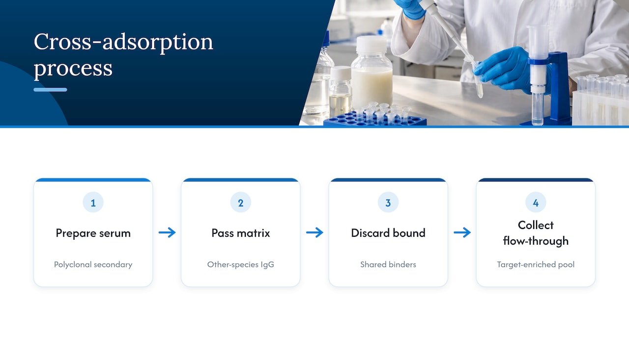
Cross-adsorption is a depletion step. The antibody preparation is passed over a matrix carrying immobilized IgG or serum proteins from other species, and the fraction that sticks is discarded. What flows through is enriched for antibodies that recognize only the intended species. The idea is old: researchers were already using cross-adsorption with western blotting to tell two closely related Rickettsia infections apart, where patient sera reacted extensively with both organisms until the shared reactivity was removed.
The result is still a polyclonal reagent. It still recognizes multiple epitopes on the target IgG, which is why it gives strong signal, but the pool is narrower than an unadsorbed one.
Why you would not want a cross-reactive secondary
In an indirect immunofluorescence workflow, a primary antibody binds the target epitope and a fluorophore-tagged secondary then binds the primary. The method amplifies signal because several secondaries can bind each primary. The flip side is that the secondary is only as selective as its own binding profile. If it also grabs the second primary on the slide, two channels end up showing the same structure, and you may read colocalization that is not there.
The same logic holds in plate-based formats. When groups develop immunoassays, non-specific binding and cross-reaction have to be prevented to avoid false results, and the detection antibody is one of the places where that is decided.
Reading the label: cross-adsorbed, highly cross-adsorbed, minimal cross-reactivity
Vendors do not use one universal vocabulary, so treat the terms below as common usage and confirm on the datasheet.
| Term on the datasheet | What it generally means | Typical use |
|---|---|---|
| Unadsorbed or “whole” | No depletion step. Broad reactivity across species. | Single-species samples, simple blots, cases with no other IgG present |
| Cross-adsorbed, pre-adsorbed | Passed over IgG or serum proteins from a defined list of species | Double labeling, tissues with mixed species contexts |
| Highly cross-adsorbed, min X | Depleted against a longer list of species | Multiplex panels with three or more host species |
| Affinity purified | Isolated on the target IgG itself. Says nothing about off-target depletion. | Often combined with cross-adsorption, but not the same thing |
| F(ab’)2 fragment | Fc region removed | Tissue with Fc receptors, live-cell or immune cell staining |
Two cautions follow from this table.
First, “affinity purified” and “cross-adsorbed” are different steps. An antibody can be affinity purified against mouse IgG and still bind rat IgG strongly.
Second, the list of species matters more than the adjective. A secondary that is cross-adsorbed against human and bovine serum proteins may be perfect for a mouse and rabbit panel on human cells and still fail when you add a rat primary. Read which species were removed, not only the headline.
When you need one, and when you do not
You probably need a cross-adsorbed secondary when
- You are multiplexing with primaries from different host species. A mouse primary plus a rabbit primary, each detected by its own species-specific secondary, is the textbook case.
- Your sample contains endogenous immunoglobulin. Mouse tissue stained with a mouse primary can pick up endogenous mouse IgG with an anti-mouse secondary. This is a different problem from cross-reactivity, and the usual fix is a blocking strategy or a different detection format, but a clean, selective secondary at least removes one more variable.
- Your blocking serum is from another species. If you block with serum from a species your secondary partly recognizes, you can create background of your own.
- You have a multiplex western blot. Two primaries, two fluorescent secondaries, two channels on one membrane. Cross-talk between secondaries looks like a band that should not exist.
- You are chasing a high signal-to-noise ratio in a low-abundance target and every source of background counts.
You may not need one when
- You are running a single primary on a single-species sample, and the secondary-only control is clean.
- You use directly conjugated primaries, where the fluorophore is attached to the primary itself and no secondary is involved.
- Your readout is a simple screening assay where a small amount of background does not move the call.
Unadsorbed secondaries are not bad reagents. They are just wider tools. The cost of using a cross-adsorbed one when you did not need it is small. The cost of skipping it when you did is a figure you cannot publish.
Cross-adsorption is not a guarantee
This is the part many datasheets understate. A published study on double immunostaining with mouse and rat primaries described an overlooked cross-reactivity in which goat anti-mouse secondary binds rat primary antibodies, and it set out methods to block that cross-species binding. Mouse and rat immunoglobulins are similar enough that depleting against one does not always clear the other, and the depletion lists on datasheets do not always include both.
The lesson is not that cross-adsorption fails. It is that the claim is always relative to a list of species and to a set of conditions, and your own tissue, fixation and antibody concentration are not on that list. Validate in your system. Our companion piece on antibody cross-reactivity, its causes, detection and fixes covers the primary side of the same problem, and our guide to antibody specificity validation shows how to build the evidence file.
How to choose secondaries for a multiplex panel
Work through the panel in this order.
- List the host species and isotype of every primary. Write them down, including the species of the tissue or cells.
- Assign each primary a secondary from a different host. Raising all secondaries in the same species, commonly donkey or goat, simplifies blocking: block with serum from that single host species, and none of your secondaries will bind it.
- Check the adsorption list for every secondary against every other species in the experiment. Primary hosts, sample species and blocking serum all count.
- Avoid pairs that are known trouble. Mouse with rat primaries is the classic one. If you can, swap one primary for a different host or use a directly conjugated version.
- Pick fluorophores with separable spectra and match them to your filter sets. Cross-reactivity and spectral bleed-through look alike on an image, so remove spectral overlap as a variable early.
- Use the same secondary concentration across channels as a starting point, then titrate.
If an isotype-specific secondary is needed, for example to discriminate two mouse monoclonals of different subclasses, expect to depend on cross-adsorption even more heavily, because subclass-specific reagents have closely related targets. Our isotype control selection checklist helps with the matching control on the primary side.
A validation workflow you can run in one afternoon
You do not need a special experiment. You need controls that cost a few extra wells or slides.
Secondary-only control
Omit the primary and incubate with secondary. This is a standard control in immunofluorescence protocols, for example the one from MIT’s microscopy core, which uses it to check for unspecific binding or cross-reaction. Any signal here is the secondary’s fault, not the primary’s.
Single-primary, all-secondaries control
For a two-color experiment, stain one slide with only primary A and then add both secondaries. Stain another with only primary B and both secondaries. Signal in the wrong channel is direct evidence of secondary cross-reactivity. Repeat for each primary in the panel.
Species-swap control
If a mouse and rat primary are in the same panel, test each secondary against the other species’ primary on a sample that does not carry the target. If you see staining, you have reproduced exactly the issue described in the double immunostaining paper, and you will want to block or change reagents.
Titration
Background rises with secondary concentration. Test a dilution series and choose the highest dilution that keeps your real signal. Buffer composition matters here, and our walkthrough on mixing and titrating antibody dilution buffer gives a practical starting point.
Document it
Record catalog number, lot, dilution, incubation time and the adsorption list. When a reviewer or a colleague asks why a panel was built that way, the answer is already in your notebook.
Common failure modes
| Symptom | Likely cause | What to try |
|---|---|---|
| Signal in channel B when only primary A is present | Secondary B binds primary A | Switch to a more highly cross-adsorbed secondary, or change one primary’s host |
| Diffuse background in secondary-only control | Secondary too concentrated, or insufficient blocking | Titrate down, extend blocking, block with serum from the secondary’s host |
| Staining in tissue from the same species as the primary | Endogenous immunoglobulin | Use a blocking step for endogenous IgG or a directly labeled primary |
| Unexpected bands on a multiplex blot | Cross-talk between secondaries, or between secondary and membrane-bound IgG | Run single-primary lanes, use highly cross-adsorbed secondaries |
| Weak signal after switching to a cross-adsorbed secondary | Narrower pool of epitope-binding antibodies | Increase concentration modestly, then recheck background |
The last row is worth remembering. Depletion removes antibodies, and the pool that is left is smaller, so you may need a somewhat different working concentration than with the unadsorbed version you used before.
Perspective: what we see on the supply side
When we talk to labs about secondary antibodies, the conversation usually starts with price or fluorophore and ends with a failed multiplex image. We think the order should be reversed. The first question is which species are in your experiment, because that decides which adsorption profile you can use. Fluorophore and format come after.
We also encourage researchers to treat the datasheet as a claim to be tested rather than a guarantee. Cross-adsorption lists describe what a vendor depleted against, not what your tissue will do. A few extra control slides at the start of a project cost less than a repeated experiment or a reviewer’s question about colocalization. As a US-based team that sources reagents globally, we would rather help you pick the right profile before you order than help you troubleshoot afterward, so ask us about the species in your panel when you contact us.
— Alina
Where our catalog fits
If you are building a panel, start from the primary side. Our antibodies category lists the primary reagents we offer, and the infectious disease antibodies page covers polyclonals and monoclonals for pathogen research. Tell us the host species and isotype of each primary, the sample species and the detection format, and we will help you work out what the secondary needs to be depleted against. For the full range, see all Mayflower Bioscience product categories. Pricing is not published on the site, so send us your panel and we will respond with a quote.
Beyond immunofluorescence: ELISA, western blot and flow
The same principles apply across formats, with different consequences.
Western blot. Multiplex fluorescent blotting is where cross-adsorption pays off quickly, since both secondaries see the same membrane and the same blocking buffer. If you are deciding between blot and plate formats for a quantitative project, our comparison of western blot and ELISA for research labs sets out how dynamic range and workflow differ.
ELISA. In sandwich formats, the detection antibody sits next to a capture antibody, often from a different species. A secondary that recognizes the capture antibody generates signal with no analyte present. Researchers developing a porcine hemoglobin assay found several factors that drive non-specific binding and cross-reaction, which is a useful reminder that reagent choice is only one of them. Plate blocking and wash stringency matter equally.
Flow cytometry and live-cell work. Fc receptors on immune cells bind the Fc region of both primary and secondary antibodies. Cross-adsorption does not solve that. Fc block or F(ab’)2 secondaries do.
ChIP and related methods. Secondary reagents are less central, but antibody specificity still decides whether you pull down the target. See our guide to choosing the right antibody for ChIP for the primary-side checks.
A short ordering checklist
Before you place an order, confirm each of these:
- Host species and isotype of every primary in the experiment
- Species of the tissue, cells or lysate
- Species of the blocking serum or protein
- Whether the secondary is cross-adsorbed, and the exact list of species depleted against
- Whether you need whole IgG or F(ab’)2 fragments
- Fluorophore or enzyme, matched to your instrument
- Lot number recorded on arrival, with a secondary-only control run on the first use
FAQ
What does cross-adsorbed mean for a secondary antibody?
It means the polyclonal secondary was passed over immobilized immunoglobulins from other species, and the fraction that bound them was removed. What remains is enriched for antibodies that recognize only the intended species. It reduces off-target binding but does not eliminate it, so controls are still needed.
What is the difference between cross-adsorbed and highly cross-adsorbed?
There is no universal definition, but highly cross-adsorbed products are generally depleted against a longer list of species than standard cross-adsorbed ones. They are aimed at multiplex panels with several host species. Always read the datasheet for the specific species list rather than relying on the label.
Do I need a cross-adsorbed secondary for a single-color experiment?
Often not, provided your secondary-only control is clean and the sample holds no immunoglobulin from other species. If your blocking serum or tissue carries IgG from another species, a cross-adsorbed secondary is a low-cost safeguard. Test with and without if the stain is critical.
Can cross-adsorbed secondaries still cross-react?
Yes. Closely related species such as mouse and rat are a known weak point, and a study on double staining with mouse and rat primaries documented goat anti-mouse secondary binding rat primaries. Run single-primary controls on every channel and, if needed, block or switch one primary’s host species.
How long can I leave a secondary antibody on?
There is no single answer, because it depends on the secondary, its concentration and the assay. Follow the datasheet or your validated protocol, and note that longer incubation and higher concentration both increase background. Titrate and run a secondary-only control before extending times.
Sources
- Serological Differentiation of Murine Typhus and Epidemic Typhus Using Cross-Adsorption and Western Blotting
- An Introduction to Performing Immunofluorescence Staining (PMC)
- Non-Specific Binding and Cross-Reaction of ELISA: A Case Study of Porcine Hemoglobin Detection
- Blocking Cross-Species Secondary Binding When Performing Double Immunostaining With Mouse and Rat Primary Antibodies
- Immunofluorescence protocol (MIT)