Western Blot vs ELISA for Research Labs: 5.3 vs 1.4 Dynamic Range
For high-throughput quantification and screening across many samples, ELISA is generally the stronger choice. When you need to confirm protein identity, check molecular weight, or detect isoforms and post-translational modifications, Western blot earns its place. Neither method wins every job, and the evidence below shows exactly where each one holds the edge.
TL;DR:
- A direct autophagy flux comparison found ELISA’s dynamic range was 5.3 versus 1.4 for Western blot, with interclass correlation scores of 0.7 versus 0.4.
- Western blot was more sensitive than ELISA in early Lyme disease samples; HSV tests showed similar sensitivity and specificity, with immunoblot resolving discrepancies.
- Colorimetric detection is simplest but least sensitive; chemiluminescent or fluorescent methods improve detection limits for both assays, while antibody quality still constrains results.
- Western blot usually takes a full day or longer, whereas ELISA often finishes in hours; ELISA plates handle 96 or 384 samples per run.
- Repeat borderline ELISA readings near the cutoff or confirm them with an orthogonal method rather than forcing a positive or negative call.
Table of Contents
- How ELISA and Western Blot Work and What Each One Reports
- Sensitivity, Dynamic Range, and Reproducibility: What the Data Shows
- Time, Throughput, and Cost: Planning Your Workflow
- When to Choose ELISA, Western Blot, or Both
- How We Support Western Blot and ELISA Workflows
- Our Take on Picking the Right Assay
- Get the Reagents and Support Your Assay Needs
- FAQ
- Sources
How ELISA and Western Blot Work and What Each One Reports
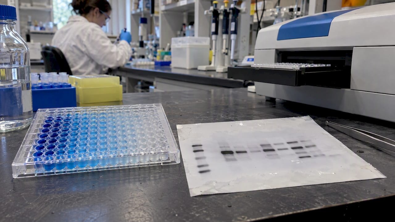
ELISA (enzyme-linked immunosorbent assay) captures a target protein on a microplate surface using antibodies, then generates a signal through an enzyme-linked detection step read as optical density. The ELISA module from the University of Colorado describes three common formats: direct, where a labeled antibody binds the antigen directly; indirect, which adds a secondary antibody step for signal amplification; and sandwich, which uses two antibodies to capture and detect the antigen with higher specificity. All three produce a readout that scales with concentration, which is what makes ELISA a quantitative workhorse.
Western blot, by contrast, separates proteins by size through gel electrophoresis, transfers them onto a membrane, then probes that membrane with antibodies to visualize bands corresponding to specific proteins. The signal appears as a band on film or a digital image rather than a single numeric value per well.
These mechanical differences drive everything downstream:
- ELISA reports a concentration value well suited to comparing many samples against each other.
- Western blot reports a band pattern that reveals size, isoform variants, and sometimes modification states that shift a protein’s apparent weight.
- Sample complexity matters differently for each: ELISA tolerates crude mixtures reasonably well because capture antibodies select the target, while Western blot’s separation step gives you visual proof you’re looking at the right protein.
- Neither method identifies a protein from sequence data alone; both depend entirely on antibody quality.
Once you see these mechanics, the rest of the comparison (sensitivity, throughput, cost) follows logically from how each assay physically works.
Sensitivity, Dynamic Range, and Reproducibility: What the Data Shows
A direct comparison of the two methods for measuring autophagy flux found that ELISA achieved a broader dynamic range of 5.3 compared with 1.4 for Western blot, along with higher reproducibility (interclass correlation of 0.7 for ELISA versus 0.4 for Western blot).
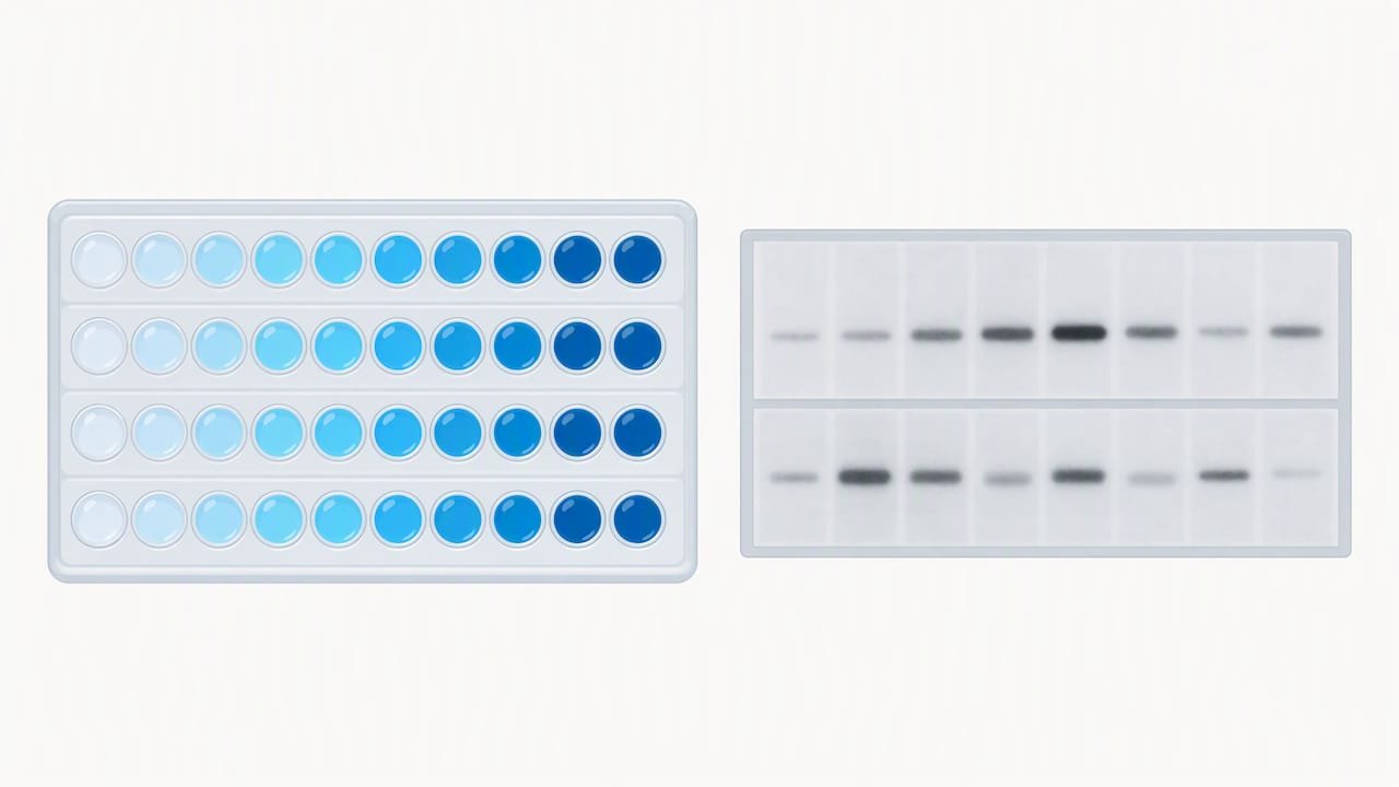
ELISA’s dynamic range advantage (5.3 vs. 1.4) and reproducibility edge (ICC 0.7 vs. 0.4) mean it can distinguish a wider spread of concentrations across replicate runs with less noise, a meaningful advantage when you’re ranking or quantifying many samples.
Diagnostic serology tells a different part of the story. Comparative evaluations of herpes simplex virus testing found that ELISA, chemiluminescent immunoassay, and immunoblot performed comparably well on sensitivity and specificity, though ELISA produced more indeterminate results in some settings, with immunoblot (Western blot) retained for discrepant resolution. In Lyme disease testing, Western blot proved more sensitive than ELISA in early-stage samples and functioned as a complementary confirmatory test rather than a first-line screen.
A few practical points explain why these patterns hold:
- Western blot variability often traces back to transfer efficiency, gel percentage selection, and antibody cross-reactivity, all of which require careful optimization.
- ELISA’s plate-based format standardizes incubation and washing steps, which reduces some sources of run-to-run variability.
- Detection chemistry changes sensitivity substantially: colorimetric detection is simplest but least sensitive, while chemiluminescent and fluorescent methods extend the limit of detection for both platforms.
- Antibody lot-to-lot consistency affects both assays, but it shows up as band inconsistency in Western blot and as shifted standard curves in ELISA.
Time, Throughput, and Cost: Planning Your Workflow
Hands-on time differs sharply between the two methods. A Western blot run, including gel casting or loading, transfer, blocking, primary and secondary antibody incubations, and imaging, typically spans a full day or longer. An ELISA plate run, once reagents and samples are prepared, often finishes in a few hours with far less hands-on manipulation per sample.
That time difference compounds at scale:
- ELISA plates process 96 or 384 samples simultaneously, which suits large cohort studies or repeated screening.
- Western blot processes a limited number of lanes per gel, so scaling up means running more gels, not just loading more samples.
- Automation exists for both, but ELISA integrates more readily with liquid handlers and plate readers for unattended runs.
Cost follows a similar pattern. Western blot costs accumulate through gels, transfer membranes, blocking reagents, and imaging consumables, often on a per-gel rather than a per-sample basis. ELISA costs center on kit price and plate reader time, which scales more predictably per sample.
Pro Tip: When turnaround time matters more than molecular detail, such as triaging a large sample set before deeper analysis, run ELISA first and reserve Western blot for the subset that needs confirmation.
When to Choose ELISA, Western Blot, or Both
Your experimental question should drive the method choice more than habit or lab tradition.
- Choose ELISA when you need to screen or quantify concentration across a large sample set, using sandwich or indirect formats depending on your target’s abundance.
- Choose Western blot when you need to confirm protein identity, verify correct molecular weight, or detect isoforms and post-translational modifications that shift band position.
- In clinical diagnostic algorithms, ELISA commonly serves as the initial screen while Western blot resolves equivocal or discrepant results, a pattern documented in HSV serology comparisons where multiplex assays showed high concordance with both methods but Western blot still resolved disputed cases.
- Consider multiplex microsphere assays or CLIA platforms when you need multiple analytes from a single small sample volume, since these platforms trade some of Western blot’s visual specificity for throughput and combined detection.
| Scenario | Recommended method | Why |
|---|---|---|
| Large cohort screening | ELISA | Higher throughput, plate automation |
| Confirming band identity or size | Western blot | Visual, size-resolved readout |
| Detecting isoforms or PTMs | Western blot | Resolves molecular weight shifts |
| Resolving discrepant ELISA results | Western blot | Functions as confirmatory step |
| Multiple analytes from limited sample | Multiplex assay | Combines detection in one run |
How We Support Western Blot and ELISA Workflows
Reliable results start with validated reagents. Our antibody catalog covers primary and secondary antibodies suited to both ELISA and Western blot applications, including infectious disease targets. For labs weighing throughput against depth of information, our AimPlex multiplex assays offer an alternative that captures multiple analytes per well.
A few resources worth bookmarking:
- Our antibody specificity guidance helps you select reagents validated for your target and species.
- Our bioassay product lines span ELISA-compatible kits and multiplex platforms for cohort-scale studies.
- Our team welcomes technical questions about reagent selection, assay design, or troubleshooting through our contact channels.
This article was prepared by Alina, drawing on published comparative studies and standard laboratory protocols.
Our Take on Picking the Right Assay
The rubric I’d give any lab: if your question is “how much,” reach for ELISA; if your question is “is this really the protein, and what shape is it in,” reach for Western blot. Borderline ELISA readings near the cutoff deserve a repeat run or orthogonal confirmation rather than a forced call, since indeterminate results cluster right at that threshold. If you’re unsure which path fits your project, reach out to our team for technical input.
— Alina
Get the Reagents and Support Your Assay Needs
Whether you’re running ELISA screens, Western blot confirmation, or scaling up to multiplex panels, reagents and kits are available to support the work, with technical support for product and protocol questions.

- Browse validated antibodies for ELISA and Western blot applications.
- Explore our bioassay and multiplex options for higher-throughput screening.
- Reagent purity matters for both platforms: inconsistent or impure peptide standards can confound results, a concern addressed in third-party verified research compounds from our partners at Purity X Peptides.
Visit our product catalog to find what your next assay run needs, or reach out to our team for a quote.
This article is general information, not a substitute for advice from a qualified doctor. Consult a qualified healthcare professional about your own circumstances before acting on anything here.
FAQ
What is a qualitative ELISA test?
A qualitative ELISA reports a positive or negative result based on whether signal crosses a defined cutoff, rather than a precise concentration value. It’s commonly used for screening applications where detecting presence or absence matters more than exact quantity.
What’s the difference between PCR and ELISA?
PCR detects and amplifies genetic material (DNA or RNA) to identify a pathogen or sequence, while ELISA detects proteins or antibodies using antibody-based capture and enzyme-linked signal readout. They answer different questions: PCR confirms genetic presence, ELISA measures protein or antibody levels.
What does a Western blot tell us?
Western blot confirms the presence of a specific protein, shows its approximate molecular weight through band position, and can reveal isoforms or modifications that shift that weight. It’s widely used as a confirmatory method when a screening test needs verification.
Is ELISA a type of Western blot?
No, they are distinct methods. ELISA is a plate-based assay that measures protein or antibody concentration in solution, while Western blot separates proteins by size on a gel before antibody detection, and the two are often used together rather than interchangeably.
Sources
- Comparisons of ELISA and Western blot assays for detection of autophagy flux
- Comparison of Western blot and enzyme-linked immunosorbent assay for diagnosis of Lyme borreliosis